Copyright
©The Author(s) 2020.
World J Orthop. Apr 18, 2020; 11(4): 232-242
Published online Apr 18, 2020. doi: 10.5312/wjo.v11.i4.232
Published online Apr 18, 2020. doi: 10.5312/wjo.v11.i4.232
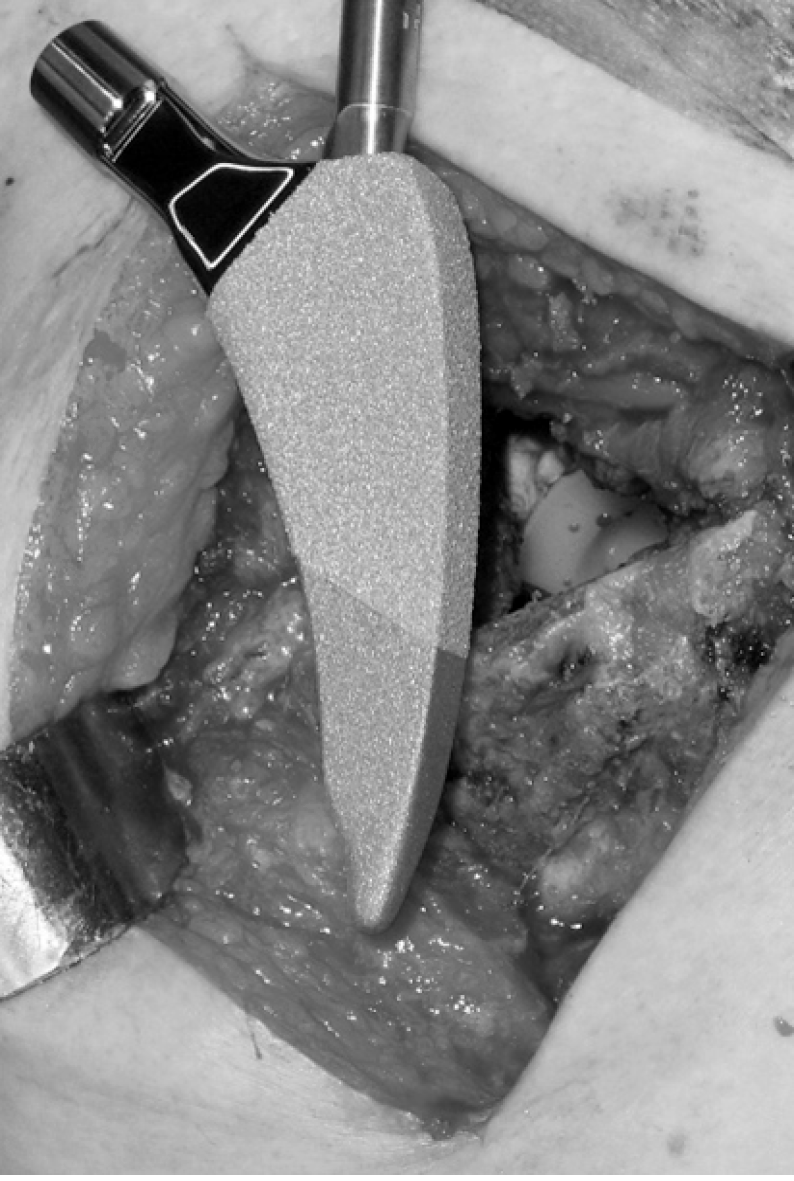
Figure 1 The MINIMA® short stem.
Figure 2 The appropriate size and orientation of the femoral component were ensured with fluoroscopy after the trial reduction in all cases.
A: Pre-operative radiograph; B: Intra-operative fluoroscopy after the trial reduction showing a varus and undersizing rasp; C: Correct orientation and sizing; D: Immediate postoperative radiograph.
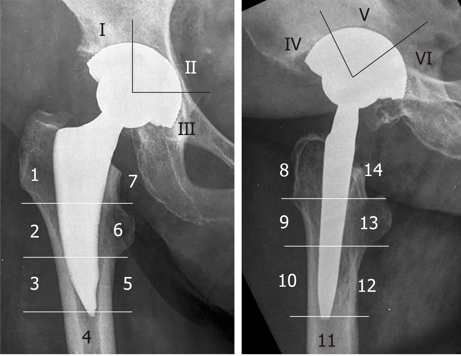
Figure 3 The modified Gruen zones.
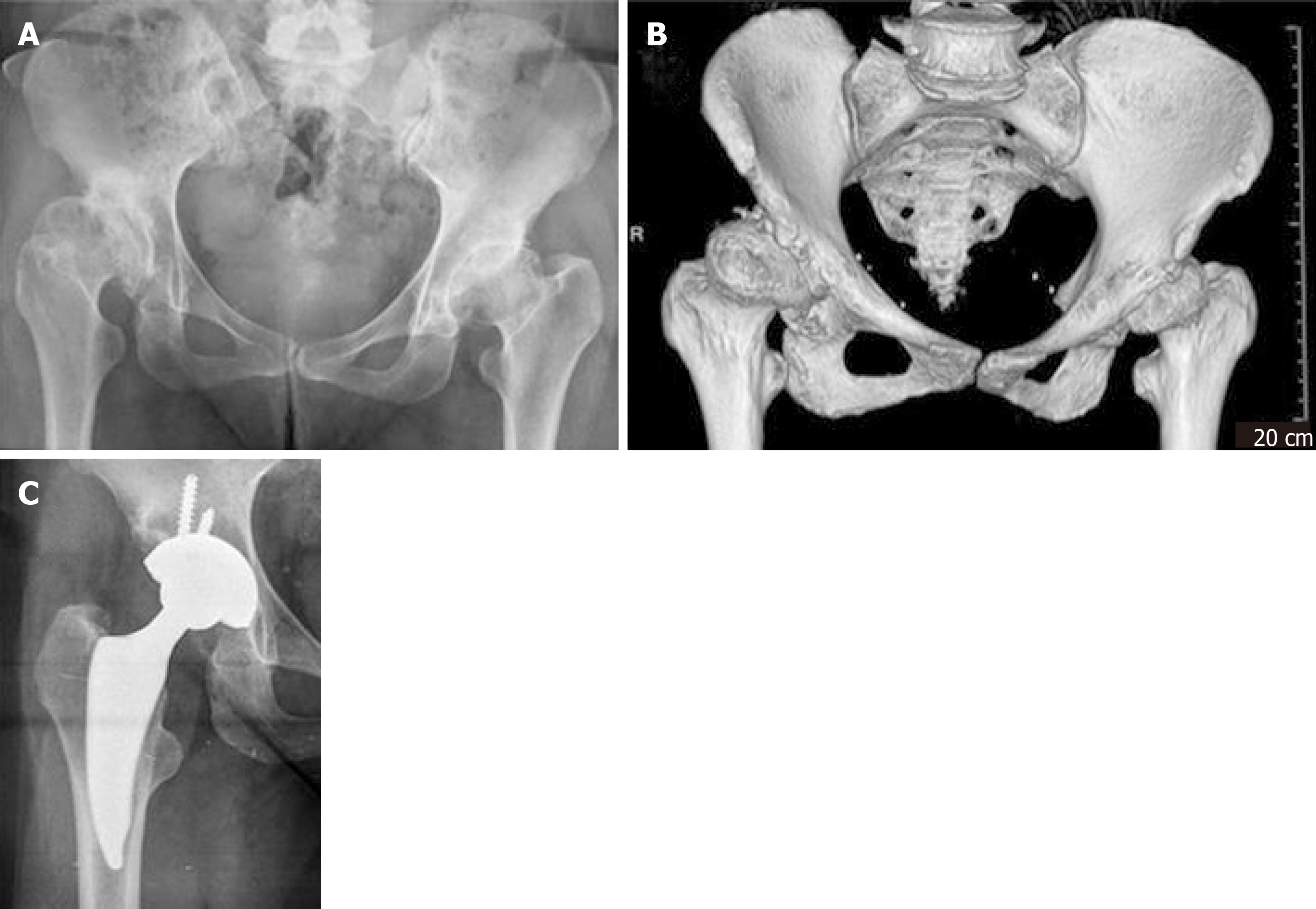
Figure 4 Hip dysplasia Hartophylakidis type II at the right side, and radiographs at 3 years postoperatively.
A, B: Hip dysplasia Hartophylakidis type II at the right side; C: Radiographs at 3 years postoperatively.
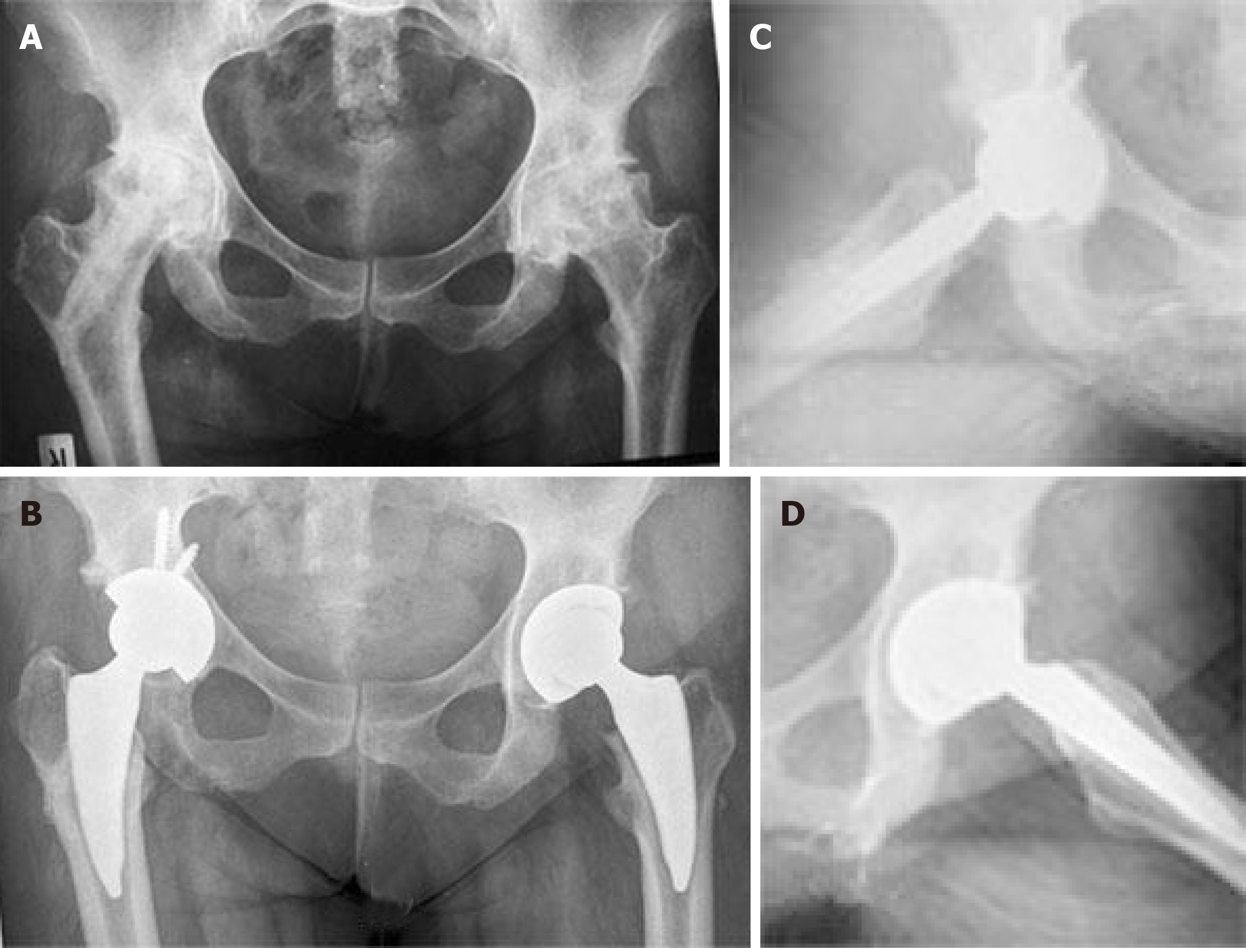
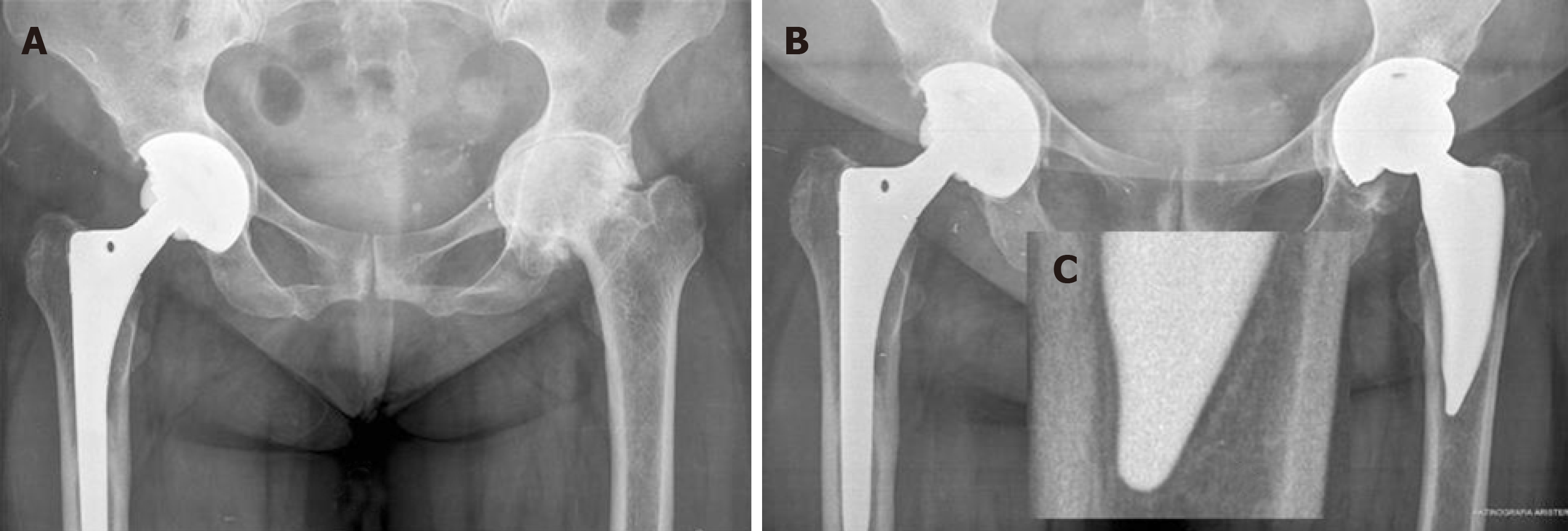
Figure 5 Bilateral osteonecrosis in a 24 years female patient suffered from lymphoma.
A: Patient treated on the right side with a vascularized fibular graft; B: Postoperative radiographs 4 years postoperatively on the right side; C: 3 years on the left side; D: Lateral radiographs.
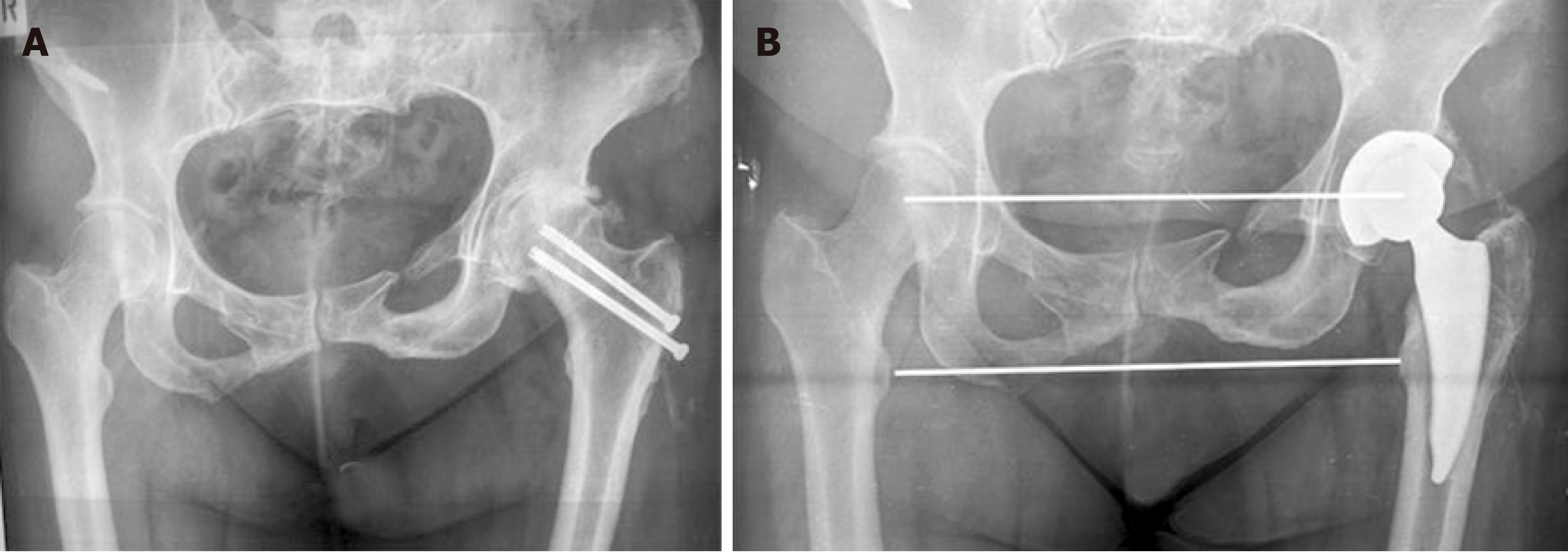
Figure 6 Ectopic ossification.
A: Posttraumatic osteoarthritis; B: Radiographs at 4 years postoperatively.
Figure 7 Distal cortical hypertrophy at modified Gruen zones 3 and 5.
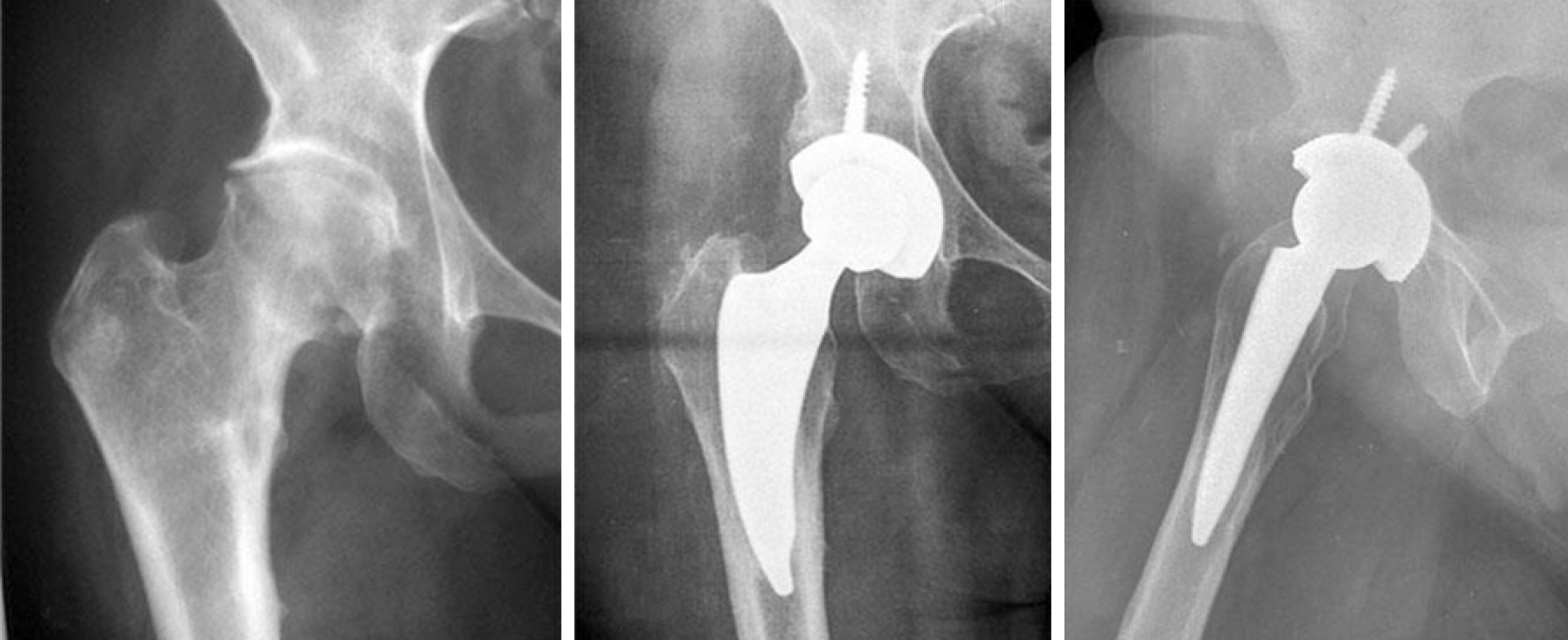
Figure 8 Reactive lines were observed in zones 3, 4 and 5.
A: Preoperative radiographs showing “protrusion acetabuli” and Dorr type C proximal femur; B: Postoperative radiographs at two years; C: Reactive lines in zones 3, 4 and 5.
- Citation: Drosos GI, Tottas S, Kougioumtzis I, Tilkeridis K, Chatzipapas C, Ververidis A. Total hip replacement using MINIMA® short stem: A short-term follow-up study. World J Orthop 2020; 11(4): 232-242
- URL: https://www.wjgnet.com/2218-5836/full/v11/i4/232.htm
- DOI: https://dx.doi.org/10.5312/wjo.v11.i4.232


