Copyright
©The Author(s) 2018.
World J Clin Oncol. Aug 13, 2018; 9(4): 60-70
Published online Aug 13, 2018. doi: 10.5306/wjco.v9.i4.60
Published online Aug 13, 2018. doi: 10.5306/wjco.v9.i4.60
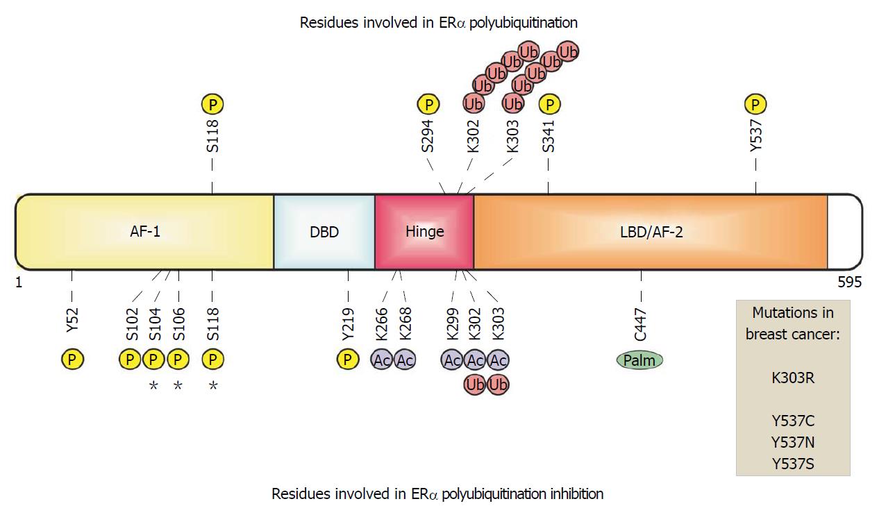
Figure 1 Estrogen receptor α in breast cancer cells.
ERα is organized in functional domains. The transactivation domains AF-1 and AF-2 recruit both coactivators and corepressors. The DNA-binding domain (DBD) recognizes and binds to estrogen response elements in enhancers or promoters. The ligand-binding domain (LBD) is recognized and activated by the 17 beta estradiol hormone. The hinge domain links LBD and DBD allowing the conformational changes of this receptor. Some residues are modified by phosphorylation, acetylation, ubiquitination and palmitoylation , which are related with ERα polyubiquitination. Sites of phosphorylation or mutations in ERα that have been identified in breast–cancer biopsy samples are indicated.
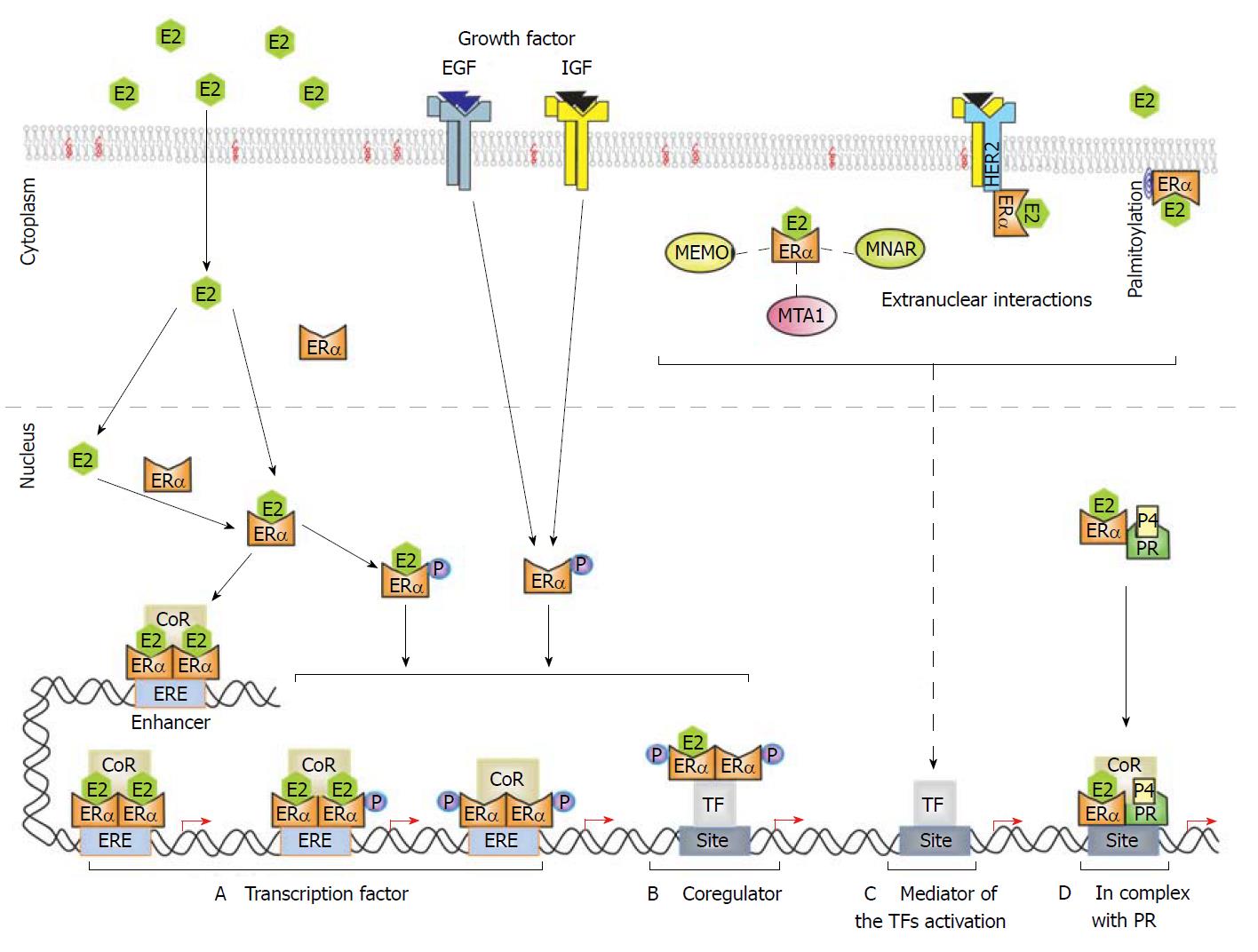
Figure 2 Nuclear and extranuclear signaling of estrogen receptor α.
E2 binds to ERα in the cytoplasm and/or nucleus. Then ERα forms homodimers that recognize the ERE sequence (AGGTCAnnnTGACCT) in target enhancers and promoters, recruiting coregulator (CoR) complexes such as coactivators to induce gene expression. ERα phosphorylation can be induced by E2 to modulate its activity as a transcription regulator. A and B: Growth factors (epidermal growth factor and insulin-like growth factor) also induce ERα phosphorylation in an E2-independent manner to promote ERα activity as a transcription factor or CoR for some transcription factors (i.e., AP-1, Sp1, and NF-κB); C: Cell membrane-associated ERα (via palmitoylation) associated with transmembranal receptors (i.e., HER2) or with cytoplasmatic proteins as (i.e., MEMO, MTA1 and MNAR). These extranuclear interactions can induce kinase–dependent signaling that could finalize in the activation of some transcription factors; D: PR can associate with ERα to coordinate the binding of ERα to the chromatin modulating the expression of specific genes.
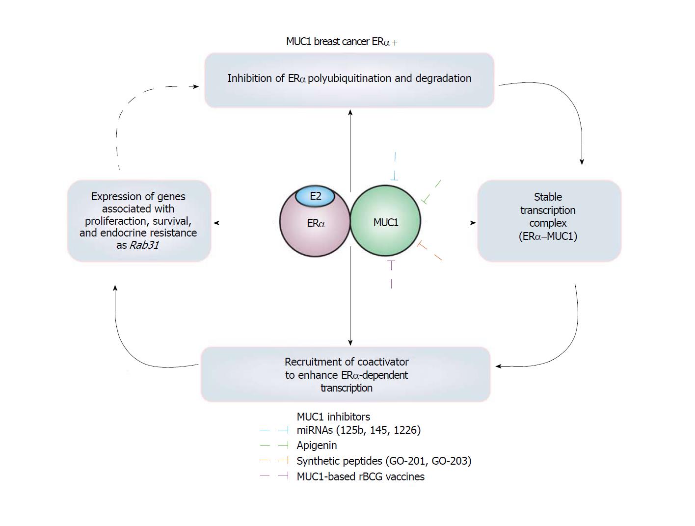
Figure 3 Mucin 1 is an estrogen receptor α polyubiquitination inhibitor protein in breast cancer cells.
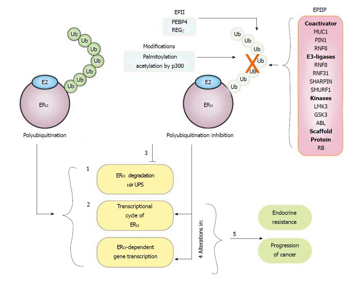
Figure 4 Mechanisms implicated in the estrogen receptor α polyubiquitination inhibition.
Half-life of estrogen receptor α protein oscillates between 3-5 h under basal condition. E2 treatment induces ERα polyubiquitination, and as result: (1) Degradation of this receptor is promoted, decreasing its protein levels starting from 1h after treatment; (2) the ERα transcriptional cycle is activated. ERα polyubiquitination inhibitor proteins (EPIP) and ERα polyubiquitination indirect inhibitors (EPII) and other modifications increased in breast cancer cells can inhibit the basal and E2-induced polyubiquitination of ERα; resulting in (3) the inhibition of its degradation and an enhancement in the ERα protein levels; (4) alterations in the transcription cycle of this receptor and the expression of its targets genes; and (5) these events seem to be associated with endocrine resistance and progression of breast cancer.
- Citation: Tecalco-Cruz AC, Ramírez-Jarquín JO. Polyubiquitination inhibition of estrogen receptor alpha and its implications in breast cancer. World J Clin Oncol 2018; 9(4): 60-70
- URL: https://www.wjgnet.com/2218-4333/full/v9/i4/60.htm
- DOI: https://dx.doi.org/10.5306/wjco.v9.i4.60


