Copyright
©The Author(s) 2015.
World J Clin Oncol. Oct 10, 2015; 6(5): 179-183
Published online Oct 10, 2015. doi: 10.5306/wjco.v6.i5.179
Published online Oct 10, 2015. doi: 10.5306/wjco.v6.i5.179
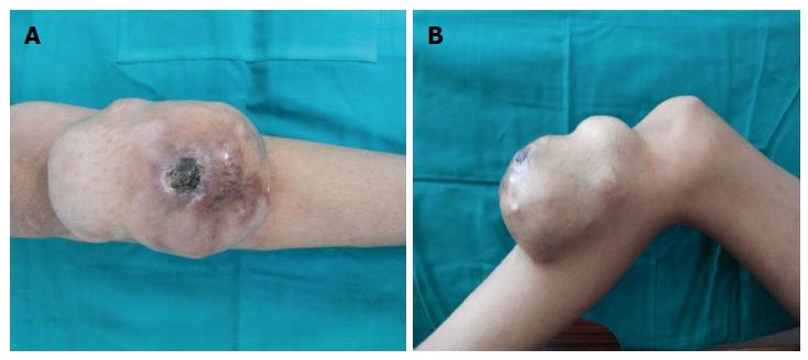
Figure 1 Preoperative clinical photographs (A and B).
Figure 2 Fine needle aspiration cytology showing loosely scattered malignant spindle cells.
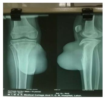
Figure 3 Preoperative X-ray.
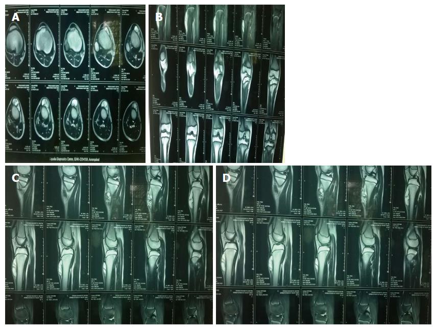
Figure 4 Magnetic resonance imaging transverse (A), coronal (B) and saggital (C, D) section.
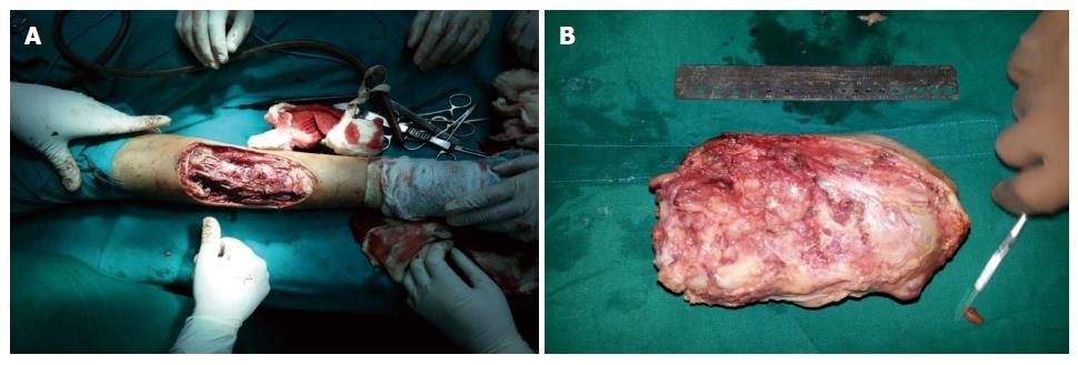
Figure 5 Intraoperative photograph showing excised mass (15 cm × 8 cm × 4 cm) (A and B).
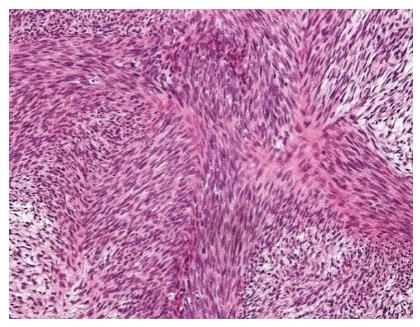
Figure 6 Malignant peripheral nerve sheath tumor on microscopy (LP 10 ×).
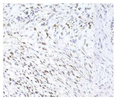
Figure 7 S-100 immunopositive tumor cells.
- Citation: Rao A, Ingle SB, Rajurkar P, Goyal V, Dokrimare N. Malignant peripheral nerve sheath tumor of proximal third tibia. World J Clin Oncol 2015; 6(5): 179-183
- URL: https://www.wjgnet.com/2218-4333/full/v6/i5/179.htm
- DOI: https://dx.doi.org/10.5306/wjco.v6.i5.179


