Copyright
©The Author(s) 2015.
World J Clin Oncol. Oct 10, 2015; 6(5): 174-178
Published online Oct 10, 2015. doi: 10.5306/wjco.v6.i5.174
Published online Oct 10, 2015. doi: 10.5306/wjco.v6.i5.174
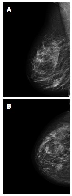
Figure 1 Mammography of the right breast shows a round lesion with smooth margins measuring 2.
6 cm in the lower inner quadrant (A and B).
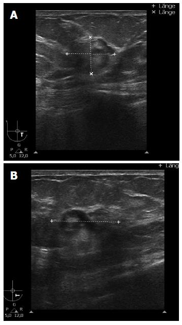
Figure 2 Breast ultrasound shows an irregular structure of complex echogenicity measuring 2.
4 cm × 2.0 cm × 1.6 cm (BI-RADS 5) (A and B).
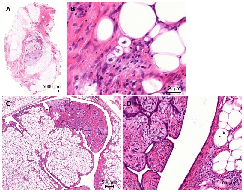
Figure 3 Lumpectomy; histopathological workup described a malignant phyllodes tumor of 21 mm diameter with a specific heterologous component identified as well differentiated liposarcoma.
A: Breast excision with centrally located phyllodes tumor (zoom × 3); B: Atypical stroma component of the phyllodes tumor including lipoblasts with multiple vacuoles (× 400); C: Intraductal phyllodes tumor with typical architecture harboring the liposarcomatous component (× 27); D: Hypercellular stroma of the phyllodes tumor showing striking atypia (left) and multivacuolated atypical lipoid cells (right) (× 200).
- Citation: Banys-Paluchowski M, Burandt E, Quaas A, Wilczak W, Geist S, Sauter G, Krawczyk N, Pietzner K, Paluchowski P. Liposarcoma of the breast arising in a malignant phyllodes tumor: A case report and review of the literature. World J Clin Oncol 2015; 6(5): 174-178
- URL: https://www.wjgnet.com/2218-4333/full/v6/i5/174.htm
- DOI: https://dx.doi.org/10.5306/wjco.v6.i5.174


