Copyright
©The Author(s) 2025.
World J Clin Oncol. Apr 24, 2025; 16(4): 103564
Published online Apr 24, 2025. doi: 10.5306/wjco.v16.i4.103564
Published online Apr 24, 2025. doi: 10.5306/wjco.v16.i4.103564
Figure 1 Contrast-enhanced computed tomography findings.
A: Jejunal space-occupying lesion; B: Thickened omentum; C: The coronal view shows the location of the tumor.
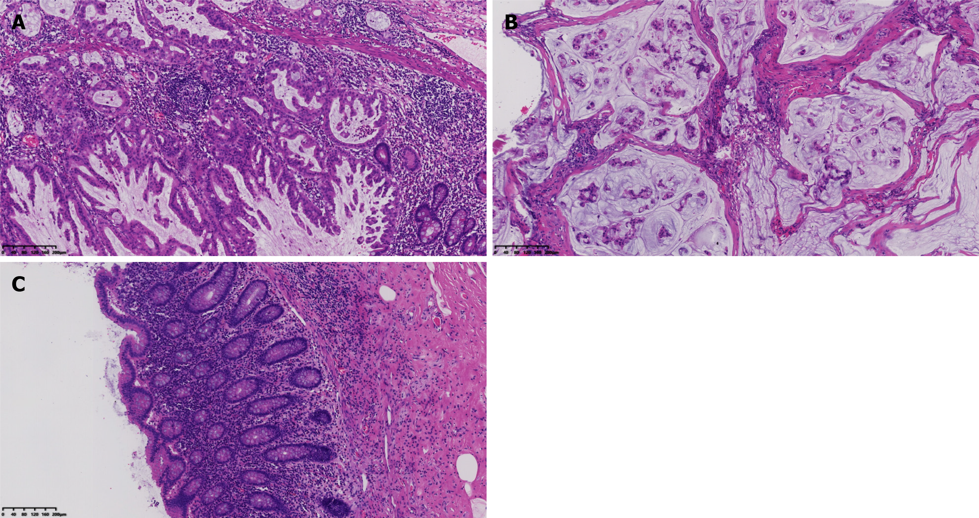
Figure 2 Pathological examinations.
A: Serrated lesions and high-grade dysplasia can be seen in the mucosa of the jejunum (× 100); B: Mucin lakes and severely dysmorphic mucus glands in the peritoneal fibrous tissue (× 100); C Normal appendix structure. Hematoxylin-eosin staining (× 100).
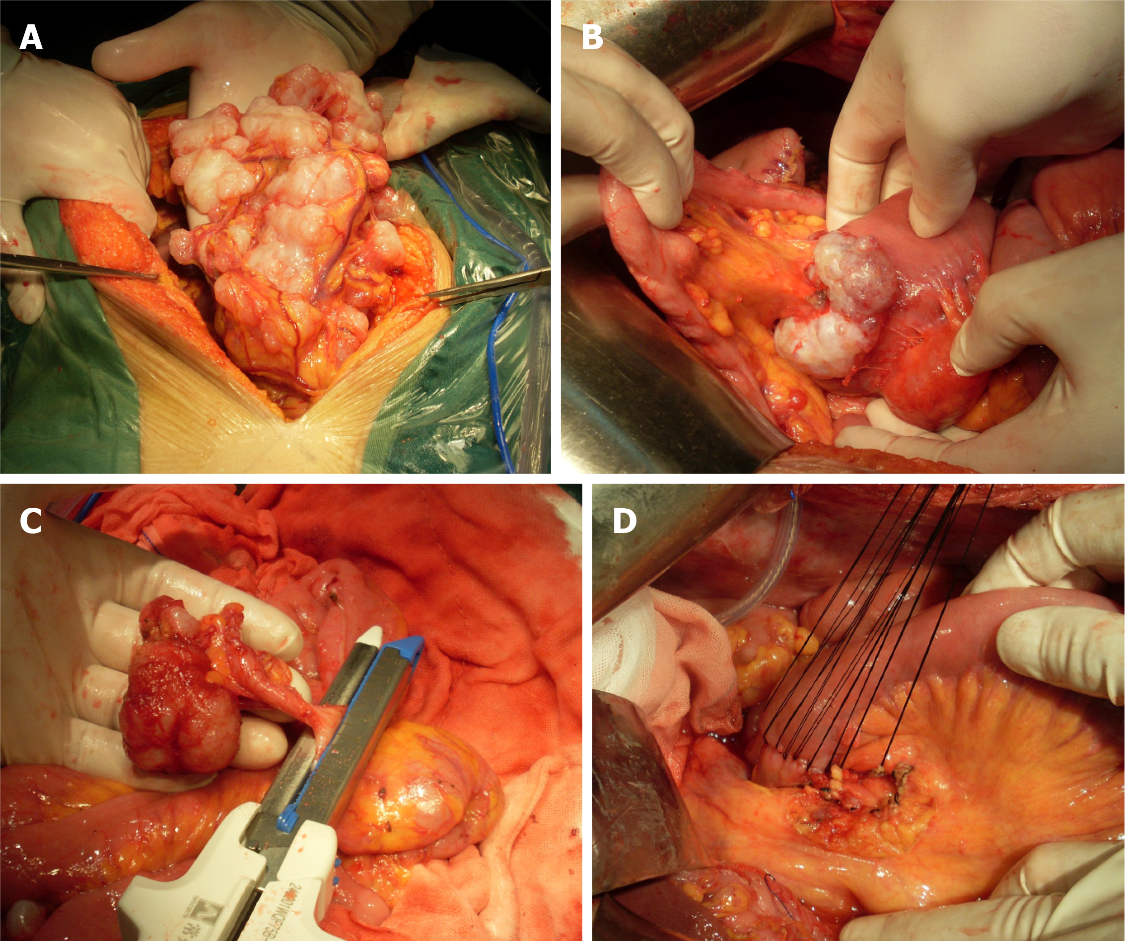
Figure 3 Surgical procedure.
A: The surface of the omentum is implanted with tumor nodules; B: A mass at the proximal jejunum; C The external appearance of the appendix is normal; D: After the jejunal mass is removed, the end-to-end anastomosis was performed.
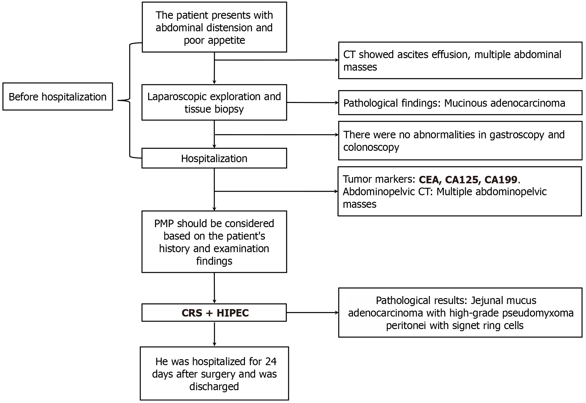
Figure 4 Diagnosis and treatment flow chart.
CT: Computed tomography; PMP: Pseudomyxoma peritonei; CEA: Carcinoembryonic antigen; CA: Carbohydrate antigen; CRS: Cytoreductive surgery; HIPEC: Hyperthermic intraperitoneal chemotherapy.
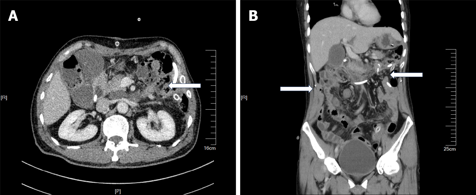
Figure 5 Postoperative contrast-enhanced computed tomography.
A: The tumour at the beginning of the jejunum disappears; B: The intra-abdominal mass were completely removed.
- Citation: Shi GJ, Wang C, Zhang P, Lu YY, Zhou HP, Ma RQ, An LB. Pseudomyxoma peritonei originating from small intestine: A case report and review of literature. World J Clin Oncol 2025; 16(4): 103564
- URL: https://www.wjgnet.com/2218-4333/full/v16/i4/103564.htm
- DOI: https://dx.doi.org/10.5306/wjco.v16.i4.103564


