Copyright
©The Author(s) 2025.
World J Clin Oncol. Mar 24, 2025; 16(3): 101909
Published online Mar 24, 2025. doi: 10.5306/wjco.v16.i3.101909
Published online Mar 24, 2025. doi: 10.5306/wjco.v16.i3.101909
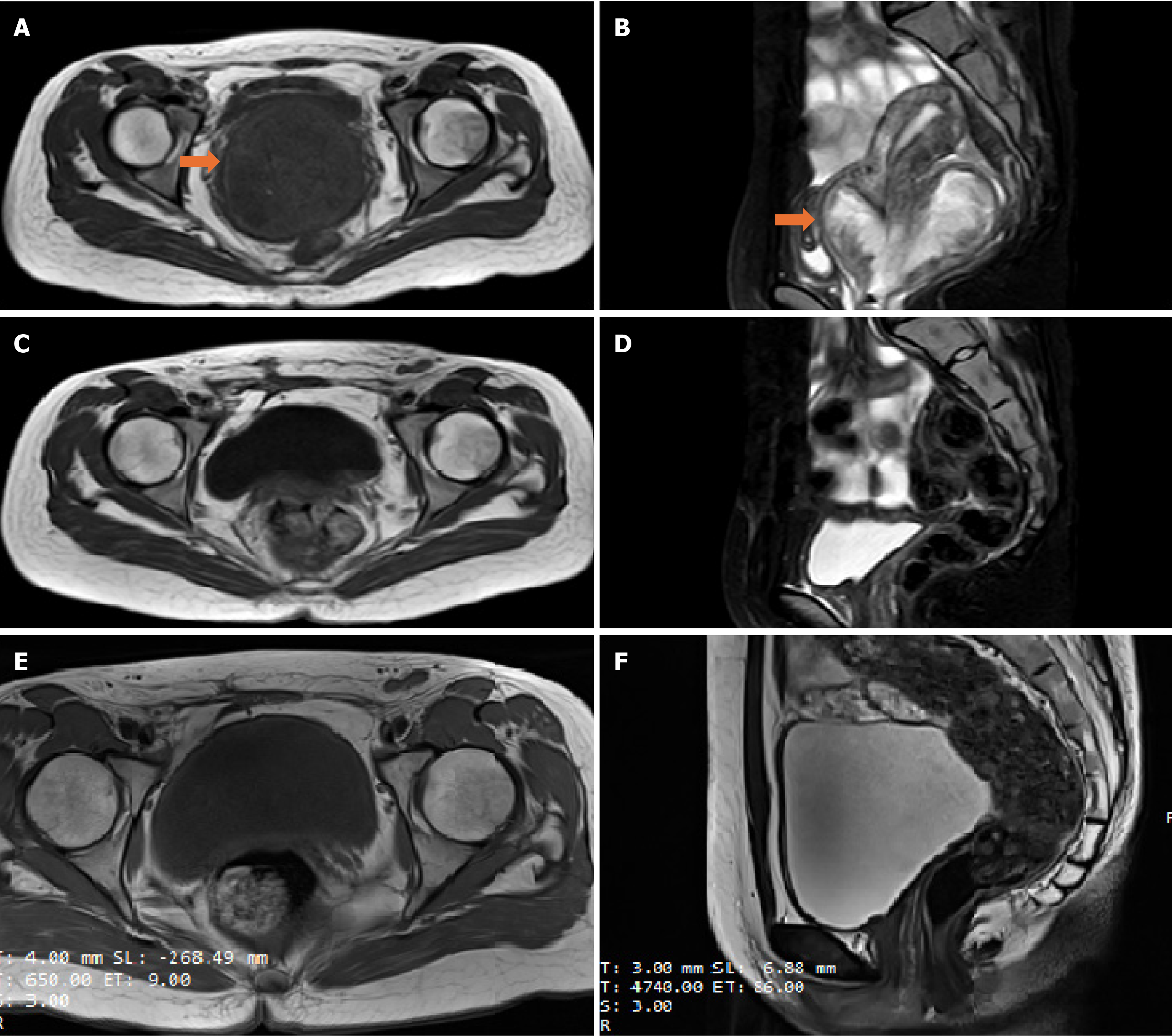
Figure 1 Patient imaging examinations.
A and B: Preoperative magnetic resonance imaging (MRI) (January 19, 2023): Cervical signal abnormalities, vaginal enlargement, and cauliflower-like mixed signal shadow, diffusion-weighted imaging showed high signal, and the maximum cross-sectional range of the lesion was 8.29 cm × 7.42 cm; C and D: Postoperative MRI (March 10, 2023): The uterus and bilateral appendages were missing, showing changes after resection, and other abnormalities were not found; E and F: The last MRI (August 26, 2024): The uterus and bilateral appendages were missing, showing changes after resection, scar shadow under right abdominal wall, and other abnormalities were not found. Orange arrow: The location of the tumor.
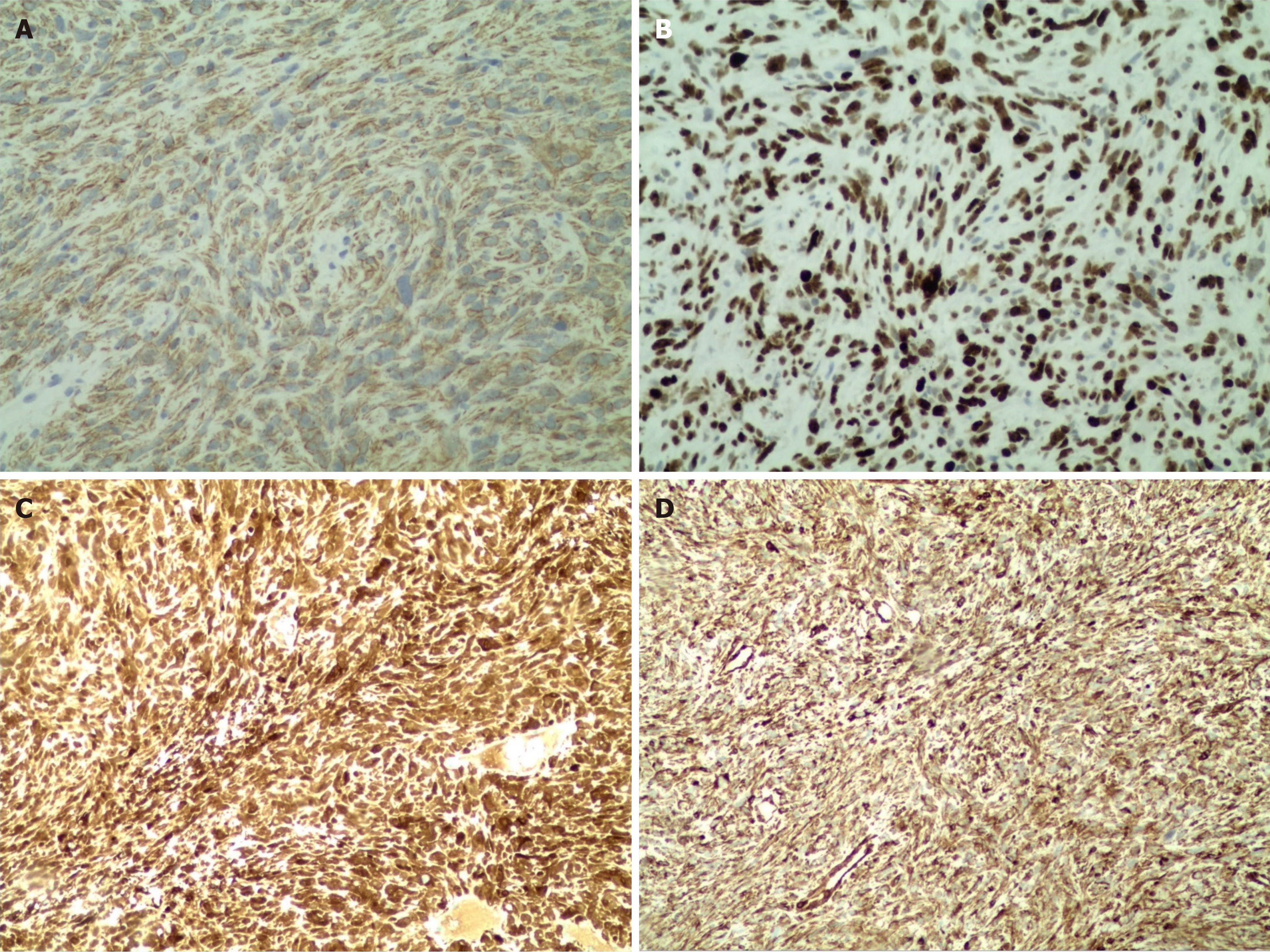
Figure 2 Immunohistochemical results.
Immunohistochemical results: CK (+), Vim (+), SATB2 (-), P16 (+), CDK4 (-), SMA (vascular +), Des (weak +), S100 (-), SMARCA4 (+), CD34 (vascular +), Ki67 (60% of hot zone), EMA (-), CD68 (histiocyte +), CD31 (vascular +), CD45RO (-), MyoD1 (-), myogenin (weak +), and SS18-SSX (-). A: Immunohistochemical results of CK; B: Immunohistochemical results of Ki67; C: Immunohistochemical results of p16; D: Immunohistochemical results of vim.
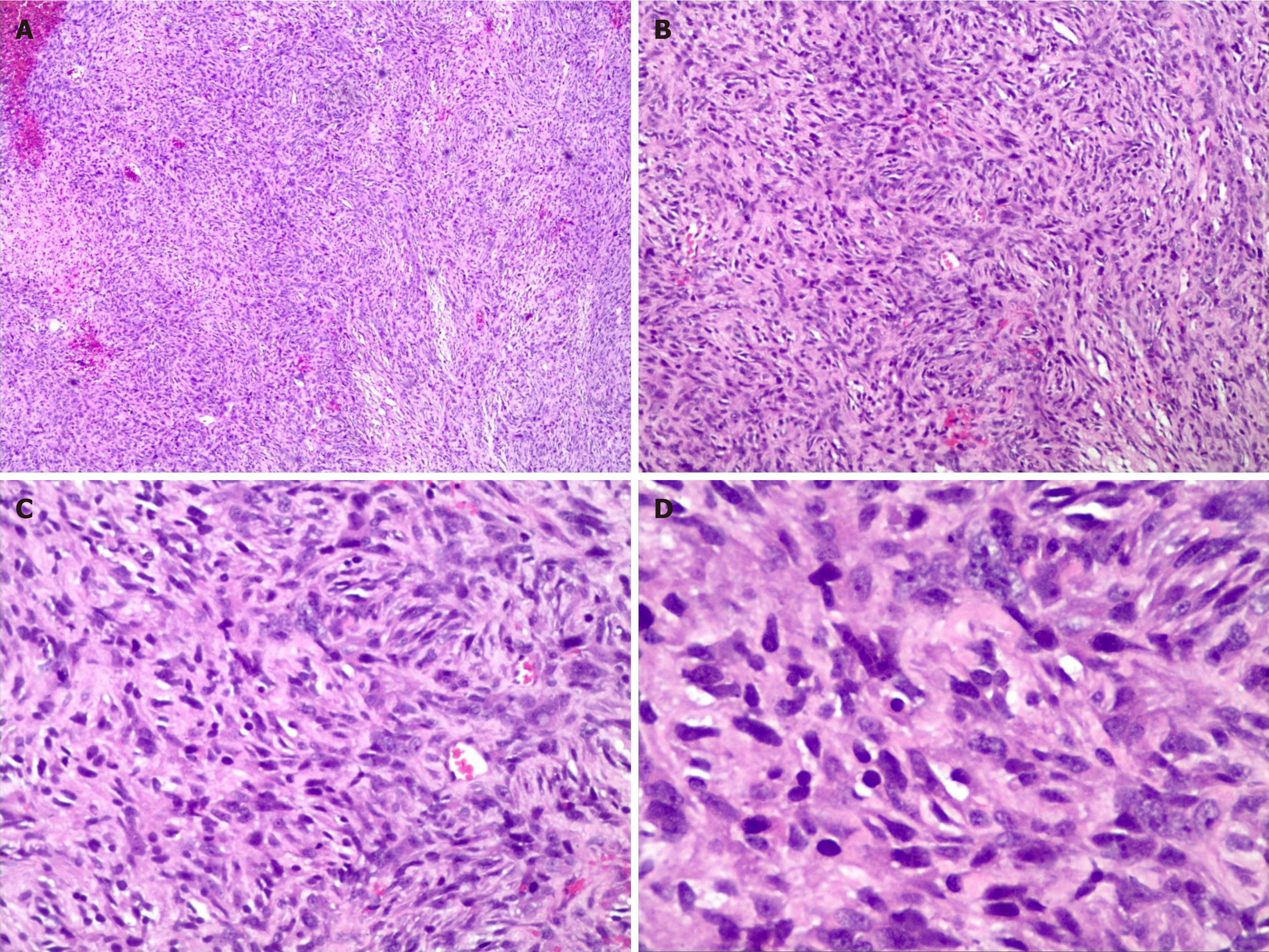
Figure 3 Hematoxylin and eosin staining at different magnifications showed that sarcoma cells could be seen.
A: Hematoxylin and eosin at 4 × 10; B: Hematoxylin and eosin at 10 × 10; C: Hematoxylin and eosin at 20 × 10; D: Hematoxylin and eosin at 40 × 10.
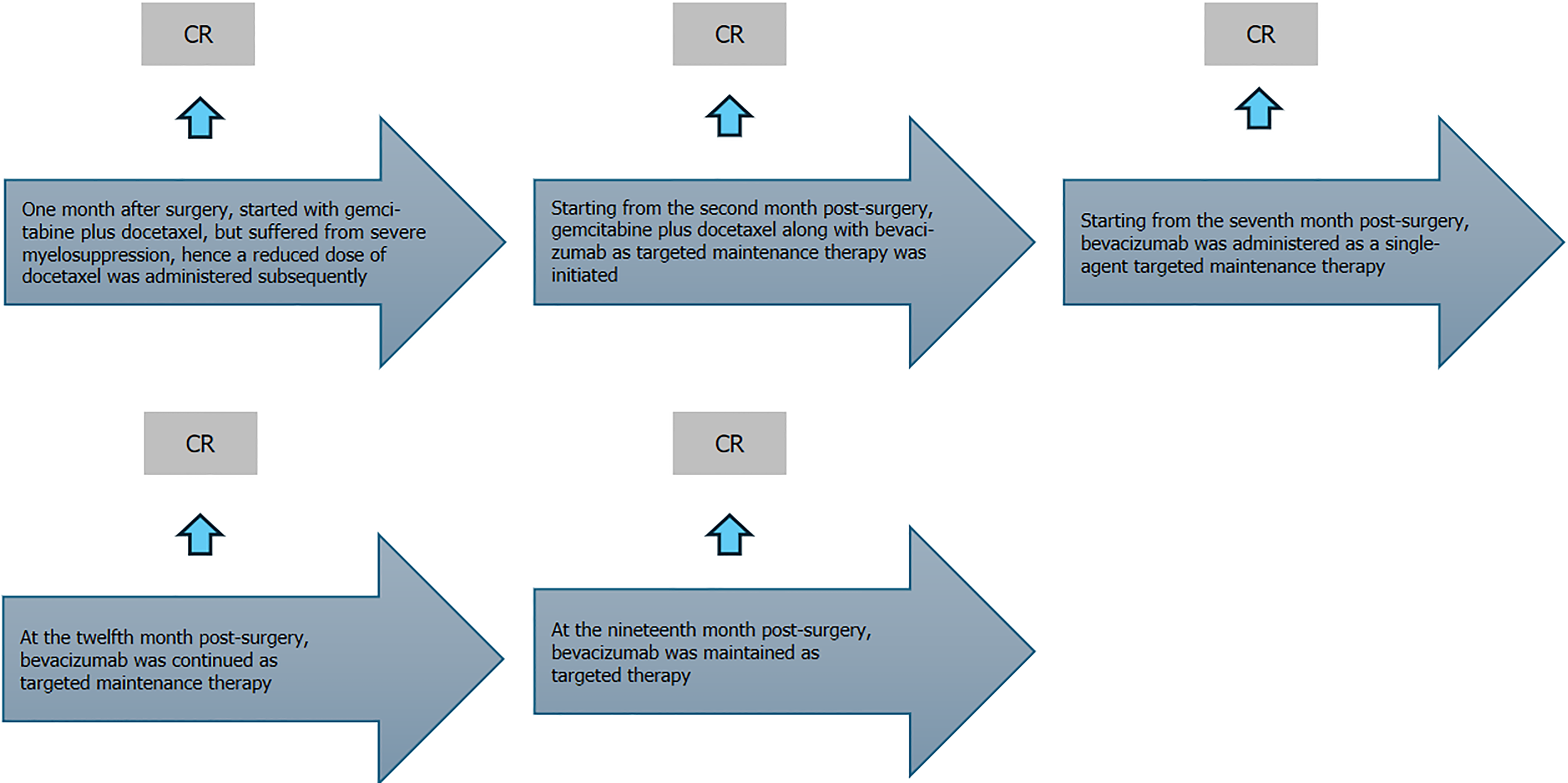
Figure 4 Evaluation of the effectiveness of the treatment process; the efficacy of the patients in the treatment and follow-up is shown in the above diagram, which is a state of continuous complete remission.
CR: Complete remission (refers to the complete disappearance of all previously detectable tumors after treatment, without any clinical and imaging evidence to prove the existence of the tumor).
- Citation: Ma HR, Zhang D, Li L, Qi L, Wang L, Li YT, Wang YR. Targeted maintenance therapy for a young woman with cervical rhabdomyosarcoma: A case report and review of literature. World J Clin Oncol 2025; 16(3): 101909
- URL: https://www.wjgnet.com/2218-4333/full/v16/i3/101909.htm
- DOI: https://dx.doi.org/10.5306/wjco.v16.i3.101909


