Copyright
©The Author(s) 2024.
World J Clin Oncol. Jun 24, 2024; 15(6): 755-764
Published online Jun 24, 2024. doi: 10.5306/wjco.v15.i6.755
Published online Jun 24, 2024. doi: 10.5306/wjco.v15.i6.755
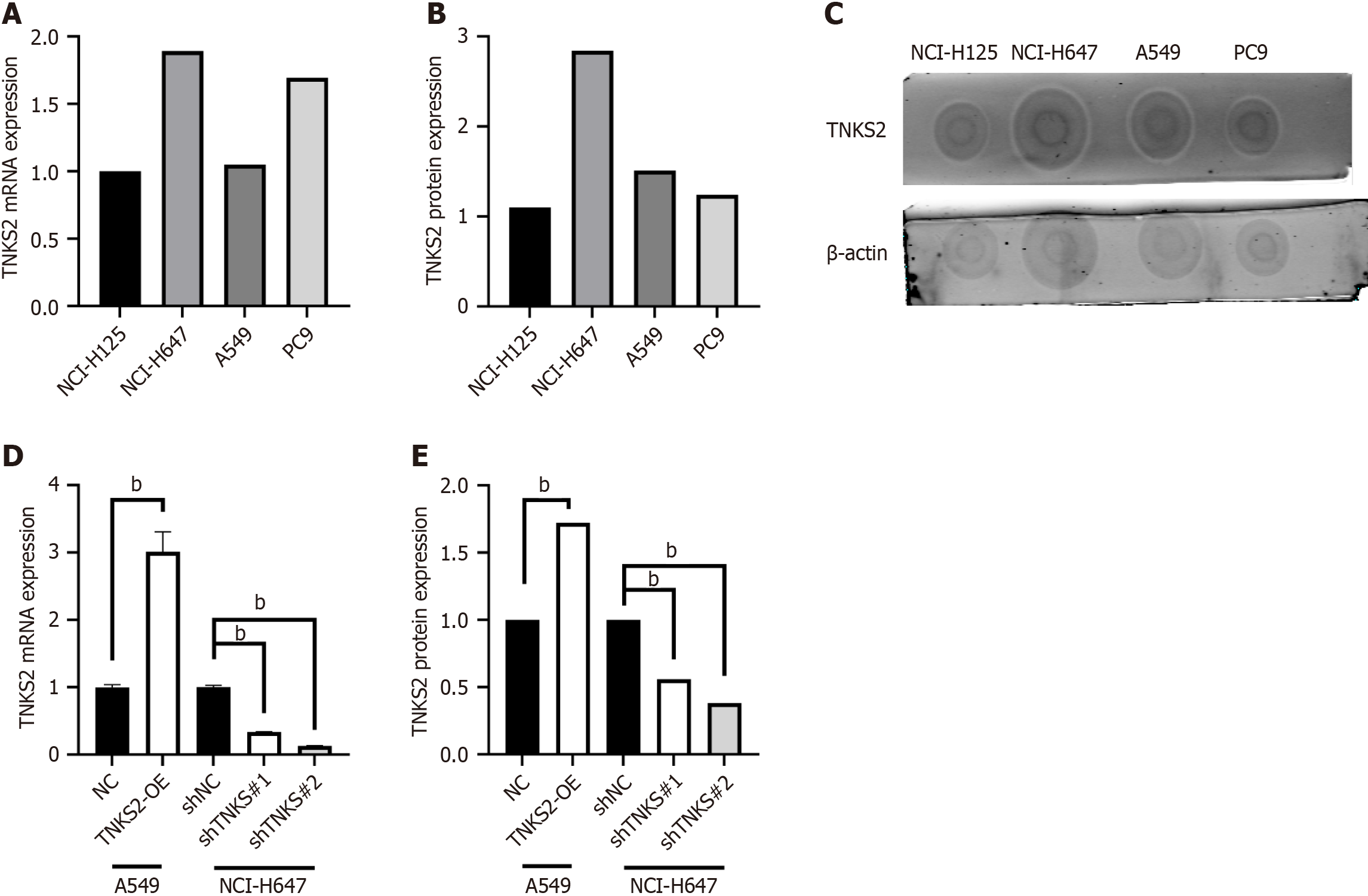
Figure 1 Successful construction of cancer cell lines with TNKS2 knockdown or overexpression.
A: Real-time reverse transcriptase-polymerase chain reaction (RT-qPCR) analysis of tankyrase 2 (TNKS2) mRNA expression in all four lung cancer cell lines; B and C: Dot-blot analysis of TNKS2 protein expression in all four lung cancer cell lines; D: Construction of stably transfected non-small cell lung cancer cell lines with TNKS2 interference and overexpression. Efficiency of TNKS2 interference or overexpression was assessed in H647 and A549 cells utilizing RT-qPCR; E: Efficiency of TNKS2 interference or overexpression was assessed in H647 and A549 cells via Western blot analysis. aP < 0.05; bP < 0.01.
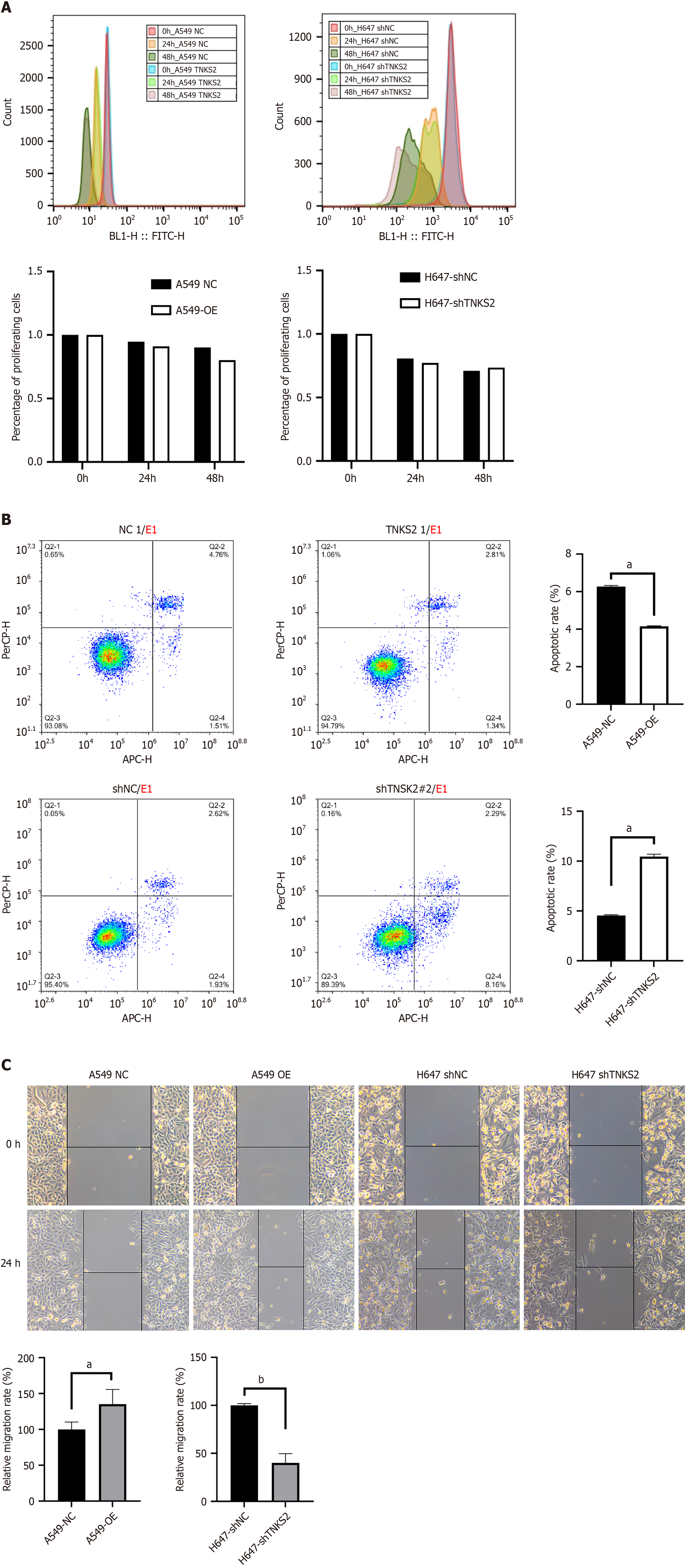
Figure 2 Effects of tankyrase 2 knockdown or overexpression on proliferation of non-small cell lung cancer cells.
A: Proliferative ability of H647 cells transfected with shTNKS2 and A549 cells with tankyrase 2 (TNKS2) overexpression; B: Apoptosis of non-small cell lung cancer cells assessed by flow cytometry. Apoptotic rate of shTNKS2-transfected H647 cells and TNKS2-overexpressing A549 cells was calculated; C and D: Migration ability of H647 cells transfected with shTNKS2 and A549 cells transfected with a TNKS2 overexpression vector at 24 h after transfection (magnification, × 100). aP < 0.05; bP < 0.01.
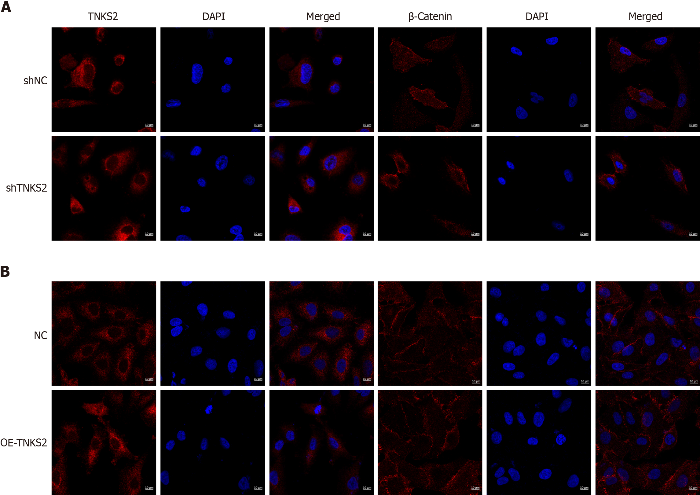
Figure 3 Immunofluorescence images obtained using confocal laser scanning microscopy.
A: Tankyrase 2 (TNKS2) and β-catenin expression in shTNKS2-transfected H647 cells; B: TNKS2-overexpressing A549 cells (magnification, × 200). Nuclei were stained blue; the target protein was stained red.
Figure 4 Expression of TNKS2/β-catenin-related proteins assessed by Western blot assay.
A: Expression levels of tankyrase 2 (TNKS2), axin, and β-catenin proteins; B: Protein expression of TNKS2-overexpressing A549 cells compared with NC-overexpressing A549 cells was analyzed by gray value; C: Protein expression of shTNKS2-transfected H647 cells compared with shNC H647 cells was analyzed by gray value. aP < 0.05; bP < 0.01.
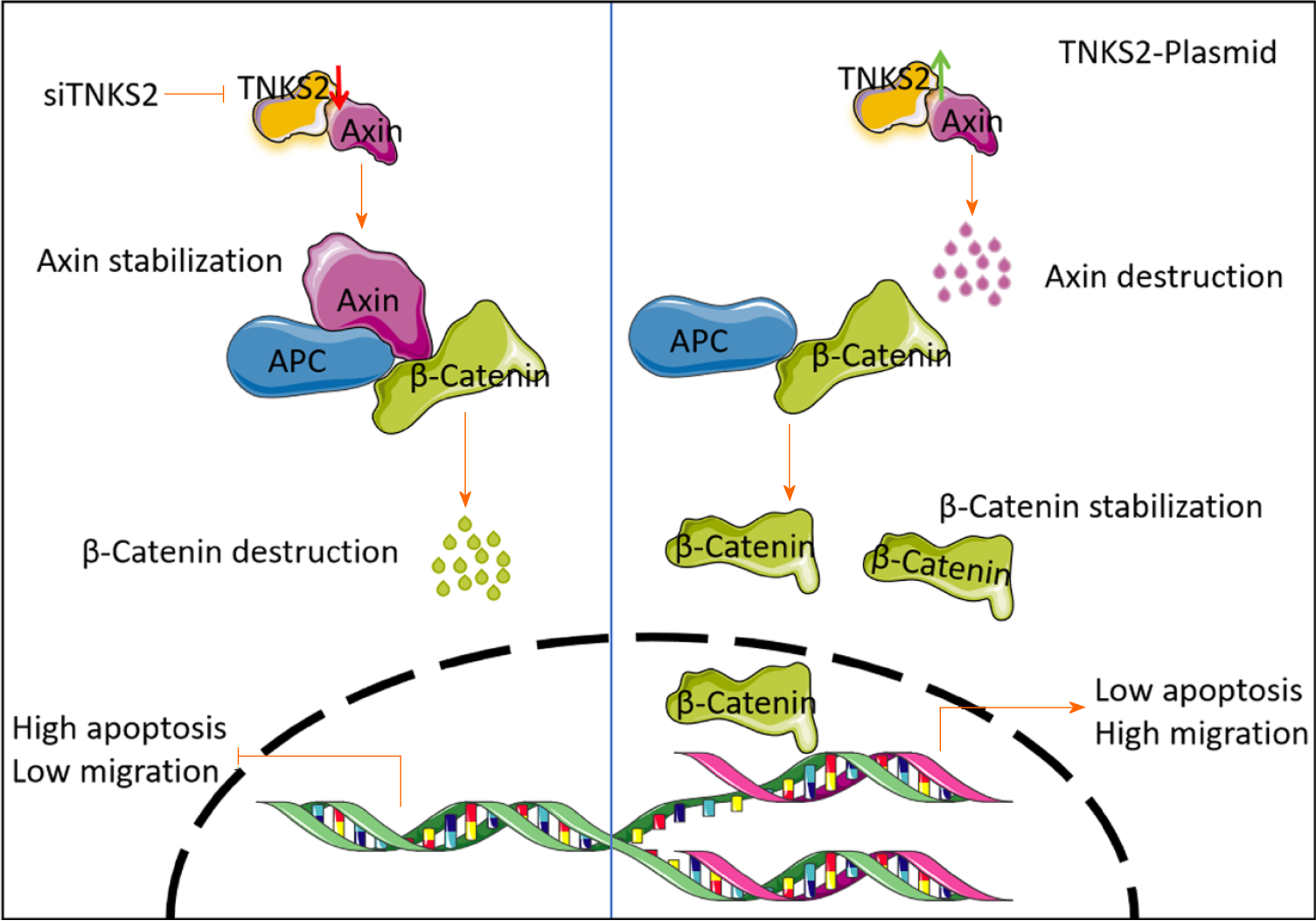
Figure 5 Tankyrase 2 overexpression promotes the malignant behavior of lung cancer cells.
Tankyrase 2 dissociates the destruction complex containing axins, thereby enhancing the stability of β-catenin. Activated β-catenin translocates into the nucleus, where it activates target genes, which contribute to the malignant behavior of lung cancer cells.
- Citation: Wang Y, Zhang YJ. Tankyrase 2 promotes lung cancer cell malignancy. World J Clin Oncol 2024; 15(6): 755-764
- URL: https://www.wjgnet.com/2218-4333/full/v15/i6/755.htm
- DOI: https://dx.doi.org/10.5306/wjco.v15.i6.755


