Copyright
©The Author(s) 2021.
World J Clin Oncol. Jan 24, 2021; 12(1): 13-30
Published online Jan 24, 2021. doi: 10.5306/wjco.v12.i1.13
Published online Jan 24, 2021. doi: 10.5306/wjco.v12.i1.13
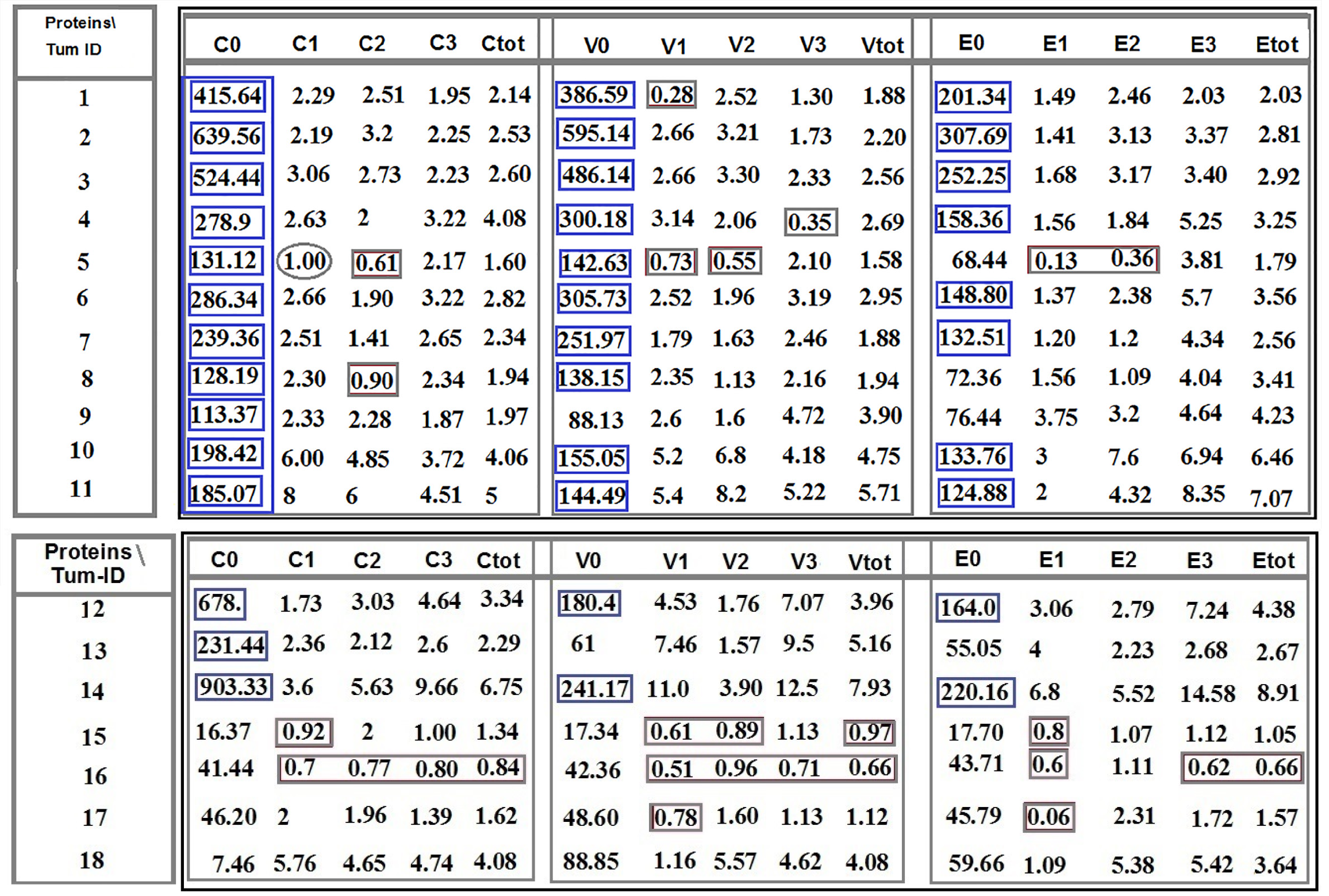
Figure 1 Ratio distribution of the cells lacking- and the classified positive-protein expression of C-C chemokine ligand 2, vascular endothelial growth factor, and epidermal growth factor between tumor cells in tumor sample to the circulated brain tumor cells.
Patients 1-11: Are affected with meningioma. Patients 12-14: Are affected with meningioma, and patients 15-18 include three metastatic carcinomas (15: Brain/lung-origin; 16 and 17: Brain breast-origin) and one with cerebellar medulloblastoma. Provided horizontal and vertical indices are related to: C0, V0, and E0: Ratio related to lack of protein expression in C-C chemokine ligand 2 (CCL2), vascular endothelial growth factor (VEGF), and epidermal growth factor (EGF), respectively; C1, V1 and E1: Low expression of CCL2, VEGF, and EGF respectively; C2, V2, and E2: Medium protein expression of CCL2, VEGF, and EGF, respectively; C3, V3, and E3: High expression of CCL2, VEGF, and EGF, respectively; Ctot, Vtot2, and Etot2: Total ratio is related to the low plus medium, plus high protein expression of CCL2, VEGF, and EGF, respectively. C: C-C chemokine ligand 2; V: Vascular endothelial growth factor; E: Epidermal growth factor.
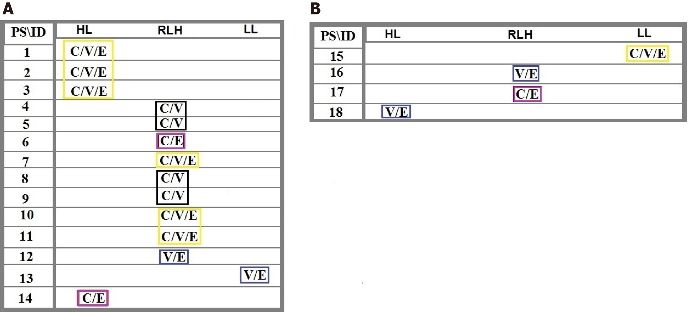
Figure 2 Ratio distribution of the cells lacking any protein expression of C-C chemokine ligand 2, vascular endothelial growth factor and epidermal growth factor between tumor cells in tumor sample to the circulated brain tumor cells in the blood.
Patients 1-14: Are affected with meningioma and patients 15-18 include 3 metastatic carcinomas (15: Brain/lung-origin; 16 and 17: Brain/breast-origin) and 1 with cerebellar medulloblastoma (CM). A: Includes patients 1-14 affected with primary meningioma; B: Includes patients 15-18 affected with the metastatic carcinoma and CM. Harmonic ratio of tumor cells to the circulated brain tumor cells are marked as specific colored quadrants related to the intensity shared classification based between different proteins. PS: Protein status; HL: Highest level; RHL: Relatively high level; LL: Low level; ID: Patients’ identification.
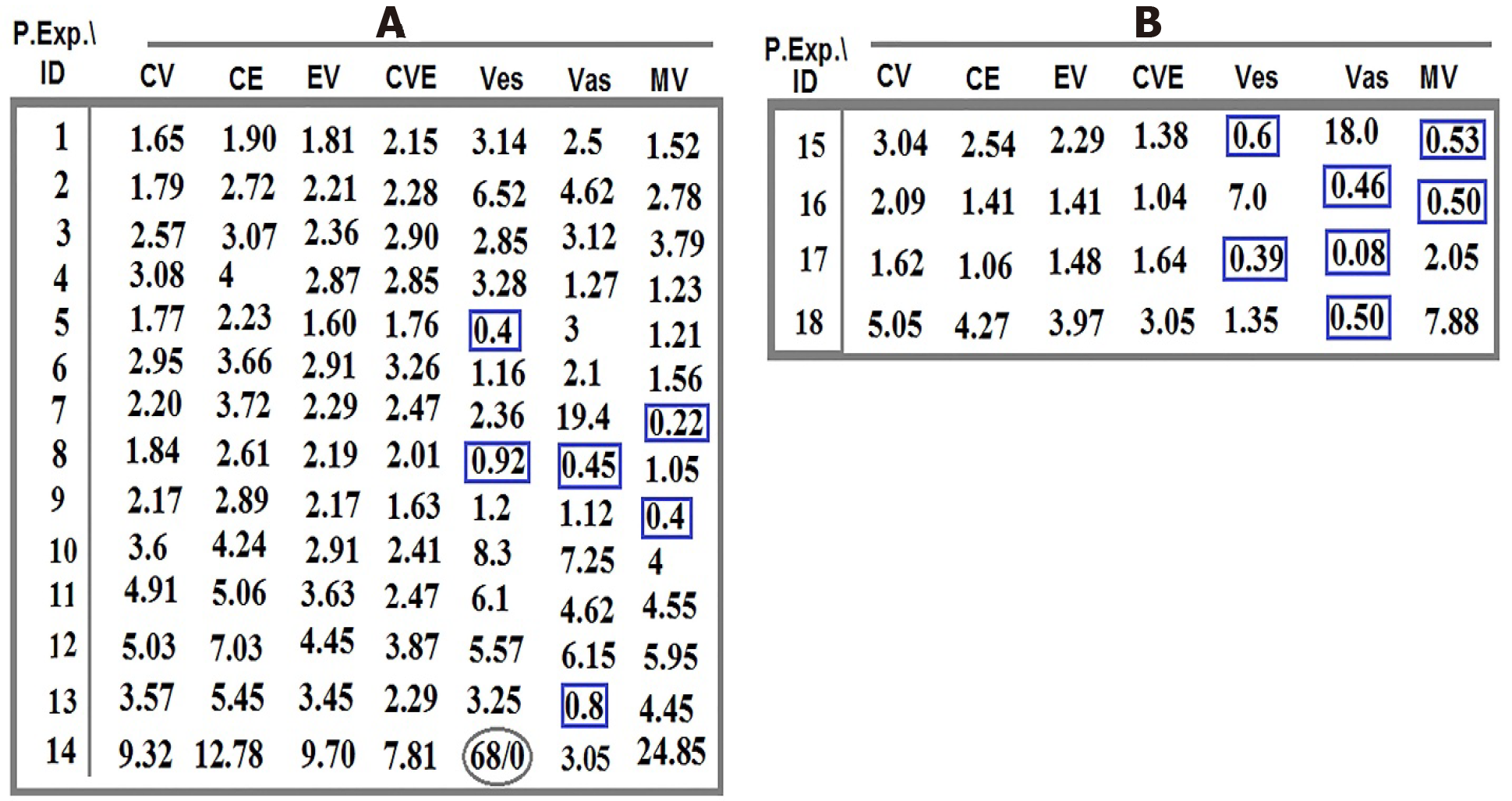
Figure 3 Ratio of co-expression of proteins between tumor cells in tumor region to circulating tumor cells and vasculature system for proteins C-C chemokine ligand 2, vascular endothelial growth factor, and epidermal growth factor in 18 patients with brain tumor.
A: The ratio of co-expression between proteins C-C chemokine ligand 2 (CCL2), vascular endothelial growth factor (VEGF), and epidermal growth factor (EGF) traced in tumor cells to the migrated tumor cells in the blood and the vascular system of patients with meningioma (pts. 1-14); B: The ratio of co-expression between proteins CCL2, VEGF, and EGF traced in tumor cells to the migrated tumor cells in the blood and the vascular system of patients with brain cancer (pts. 15-18). The highlighted quadrants with different colors indicate the harmonic evolutionary altered intensity of related proteins in common amongst different patients. Pr. Exp.: Protein expression; ID: Patients’ identification; C: C-C chemokine ligand 2; V: Vascular endothelial growth factor; E: Epidermal growth factor; Ves: Vesicles; Vas: Vasculature; MV: Microvesicles (angiogenesis).
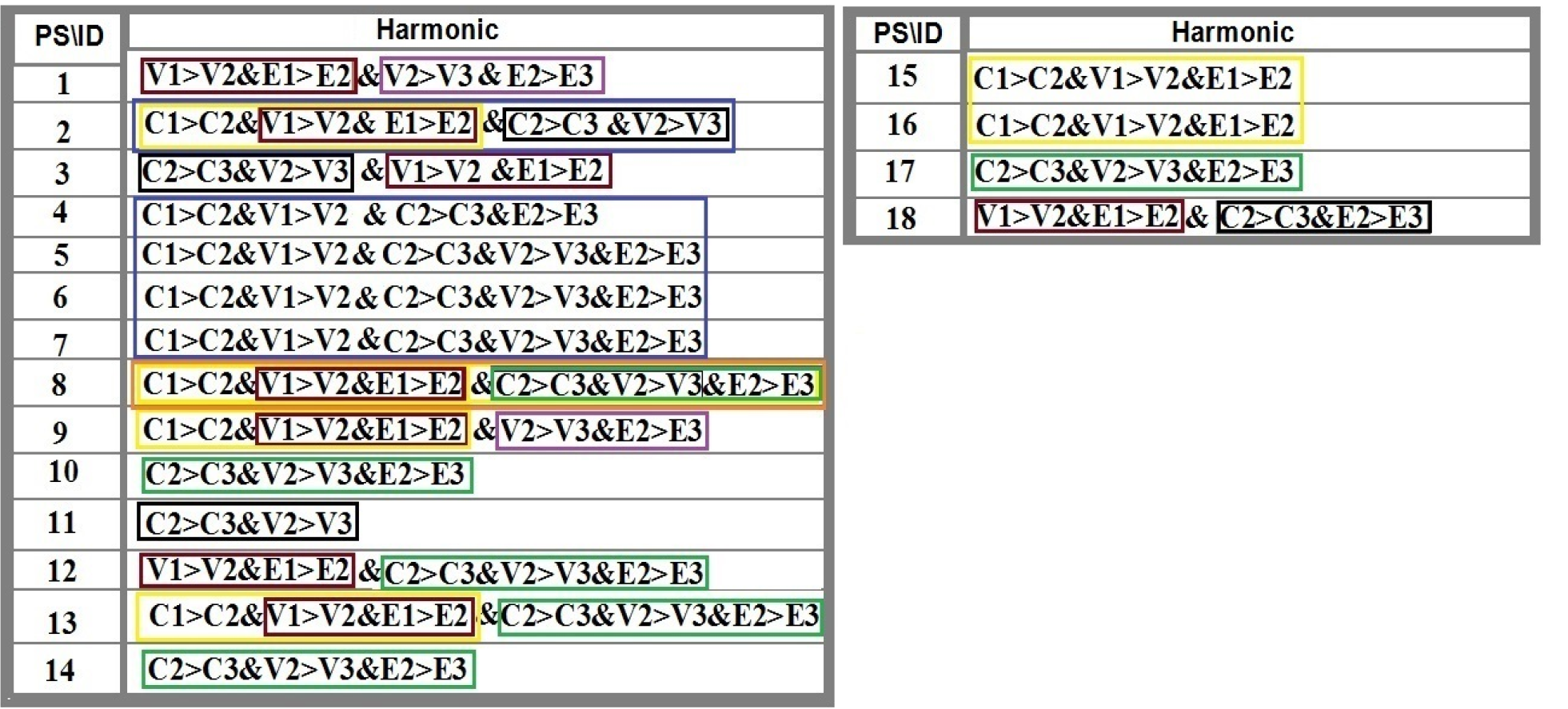
Figure 4 The harmonic ratio of tumor cells with different degree of expression intensity for protein C-C chemokine ligand 2, vascular endothelial growth factor, and epidermal growth factor to the circulating tumor cells in patients with brain tumors.
C-C chemokine ligand 2 (CCL2), vascular endothelial growth factor (VEGF), and epidermal growth factor (EGF) are conjugated with fluorescein isothiocyanate, R-phycoerythrin, and phycoerythrin–indodicarbocyanine, respectively. The highlighted quadrants with different colors indicate the harmonic evolutionary altered intensity including increased and/or decreased ratio value of the related proteins in common between different patients or within different proteins. The altered ratio is based on the provided data in Figure 1; Numbers 1 (high), 2 (medium) and 3 (low) are reflective of expression intensity of proteins CCL, VEGF and EGF. C: C-C chemokine ligand 2; V: Vascular endothelial growth factor; E: Epidermal growth factor.
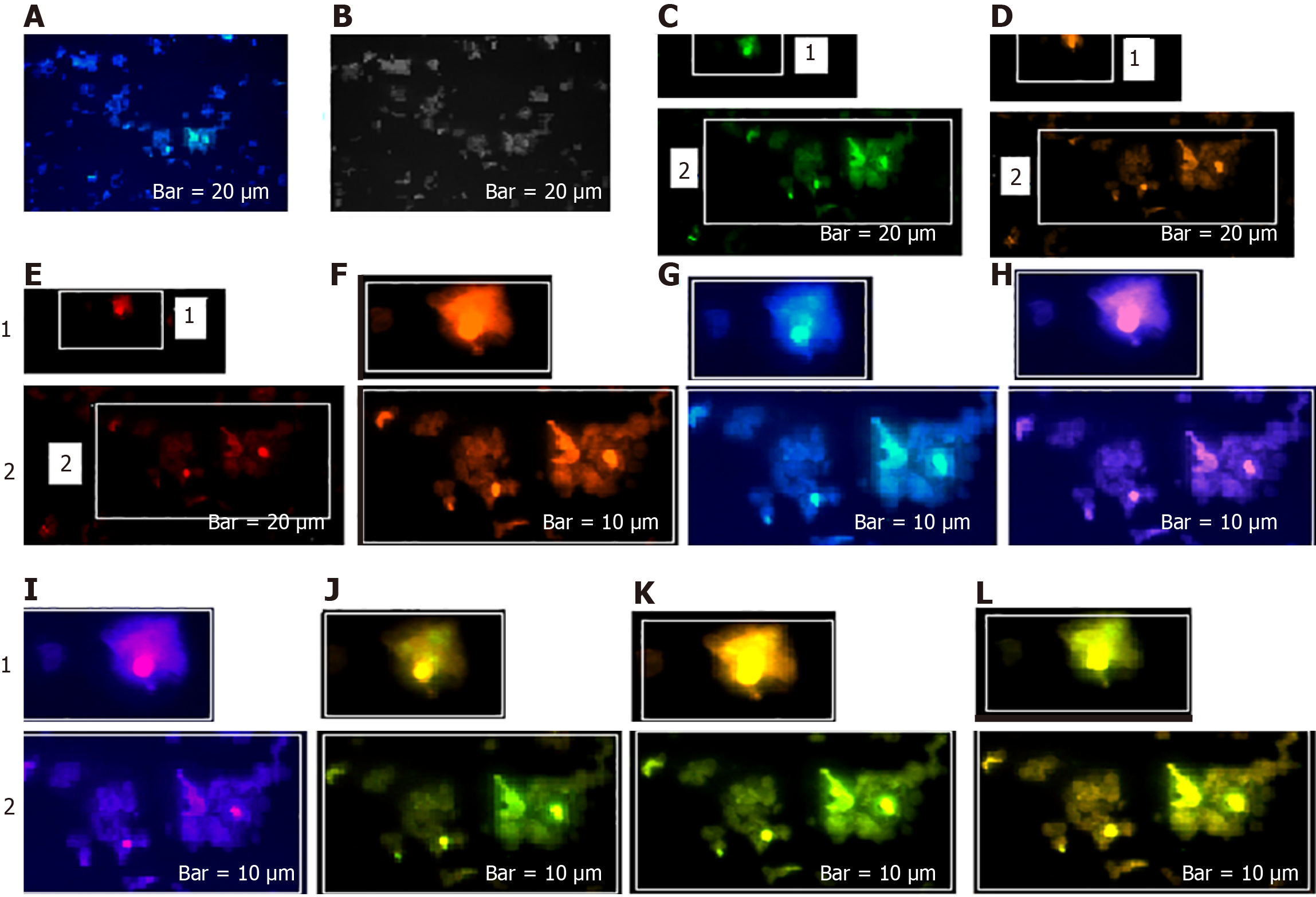
Figure 5 Mode of protein expression and co-expression of C-C chemokine ligand 2, vascular endothelial growth factor, and epidermal growth factor in circulating tumor cells of a patient with meningioma.
C-C chemokine ligand 2 (CCL2), vascular endothelial growth factor (VEGF) and epidermal growth factor (EGF) are conjugated with fluorescein isothiocyanate, R-phycoerythrin, and phycoerythrin–indodicarbocyanine, respectively, in patient number 1. A: Merged of 4′,6-diamidino-2-phenylindole/CCL2; B: Phase contrast; C: Same cells with CCL2; D: Cells with VEGF; E: Cells with EGF; F: Merged/cropped cells with VEGF/EGF; G: Merged of 4′,6-diamidino-2-phenylindole /CCL2; H: Merged cells with dai/VEGF; I: Merged cells with dai/EGF; J: Cropped/merged cells with CCL2/VEGF; K: Cropped/merged cells with CCL2/EGF; L: Cropped/merged cells with CCL2/VEGF/EGF.
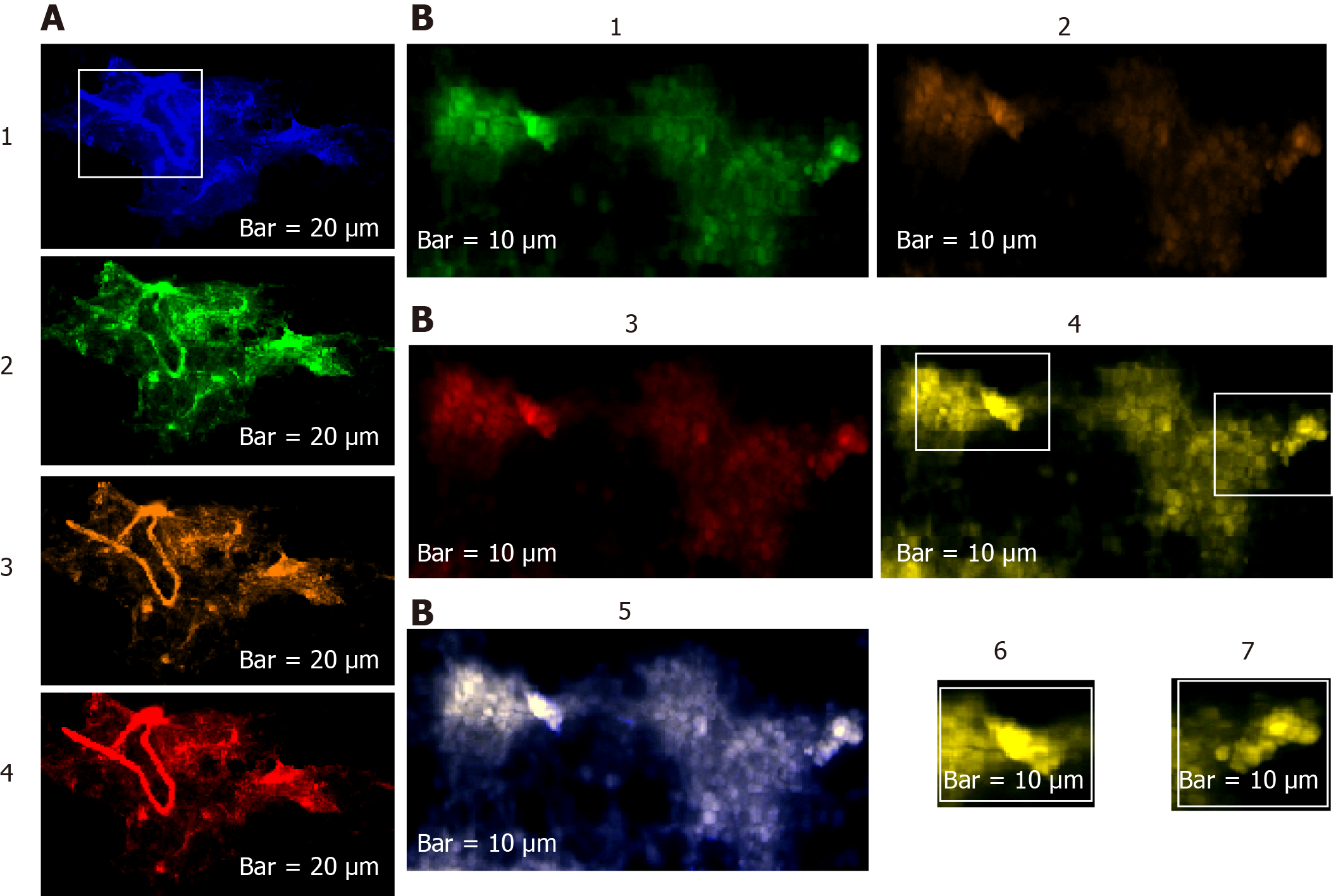
Figure 6 Status of protein expression of C-C chemokine ligand 2, vascular endothelial growth factor, and epidermal growth factor in the tumor and circulating tumor cells of patient 4 affected with meningioma.
C-C chemokine ligand 2 (CCL2), vascular endothelial growth factor (VEGF), and epidermal growth factor (EGF) are conjugated with fluorescein isothiocyanate, R-phycoerythrin, and phycoerythrin–indodicarbocyanine, respectively, in patient number 2. A1-4: 4′,6-Diamidino-2-phenylindole, CCL2, VEGF, EGF; B1-5: CCL2, VEGF, EGF, co-expression of CCL2/VEGF/EGF, co-expression of 4′,6-diamidino-2-phenylindole/CCL2/VEGF/EGF, Selected cropping area of image B4, is reflective of diverse co-expression between these three proteins from image B4.
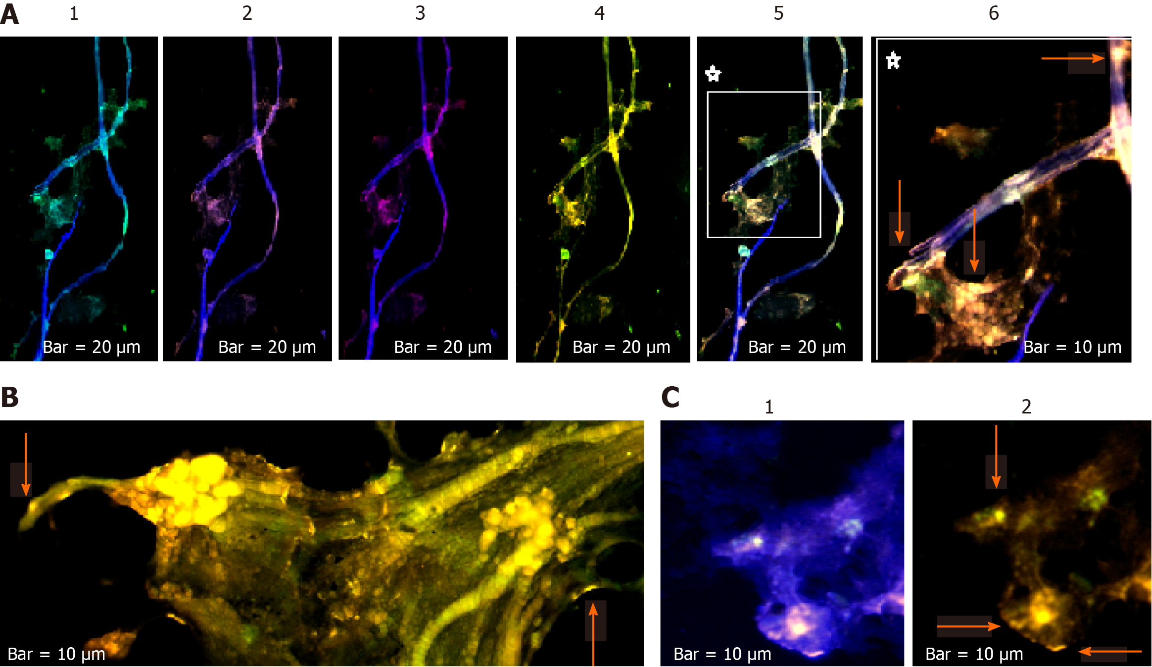
Figure 7 Protein expression status of C-C chemokine ligand 2/vascular endothelial growth factor/epidermal growth factor of two cell population from tumor sample and circulating tumor cells in patient 11 affected with meningioma.
C-C chemokine ligand 2 (CCL2), vascular endothelial growth factor (VEGF), and epidermal growth factor (EGF) are conjugated with fluorescein isothiocyanate, R-phycoerythrin, and phycoerythrin–indodicarbocyanine, respectively, in patient number 3. A: Vascular images accompanied by the partial tumor section [1: Merged of 4′,6-diamidino-2-phenylindole (DAPI)/CCL2; 2: Merged of DAPI/VEGF; 3: Merged of DAPI/ EGF; 4: Co-expression of CCL2/EGF/VEGF; 5: Co-expression of DAPI/CCL2/VEGF/EGF; 6: A cropped section of 5]; B: Image of a tumor section reflecting of the Co-expression of CCL2/VEGF/EGF; C [1:DAPI/CCL2/VEGF/EGF; 2:CCL2/VEGF/EGF]: Illustrates the CTCs in the blood sample of the same patient. The arrows are indicative of expression status in tumor cells, in vasculature, and the microvesicles.
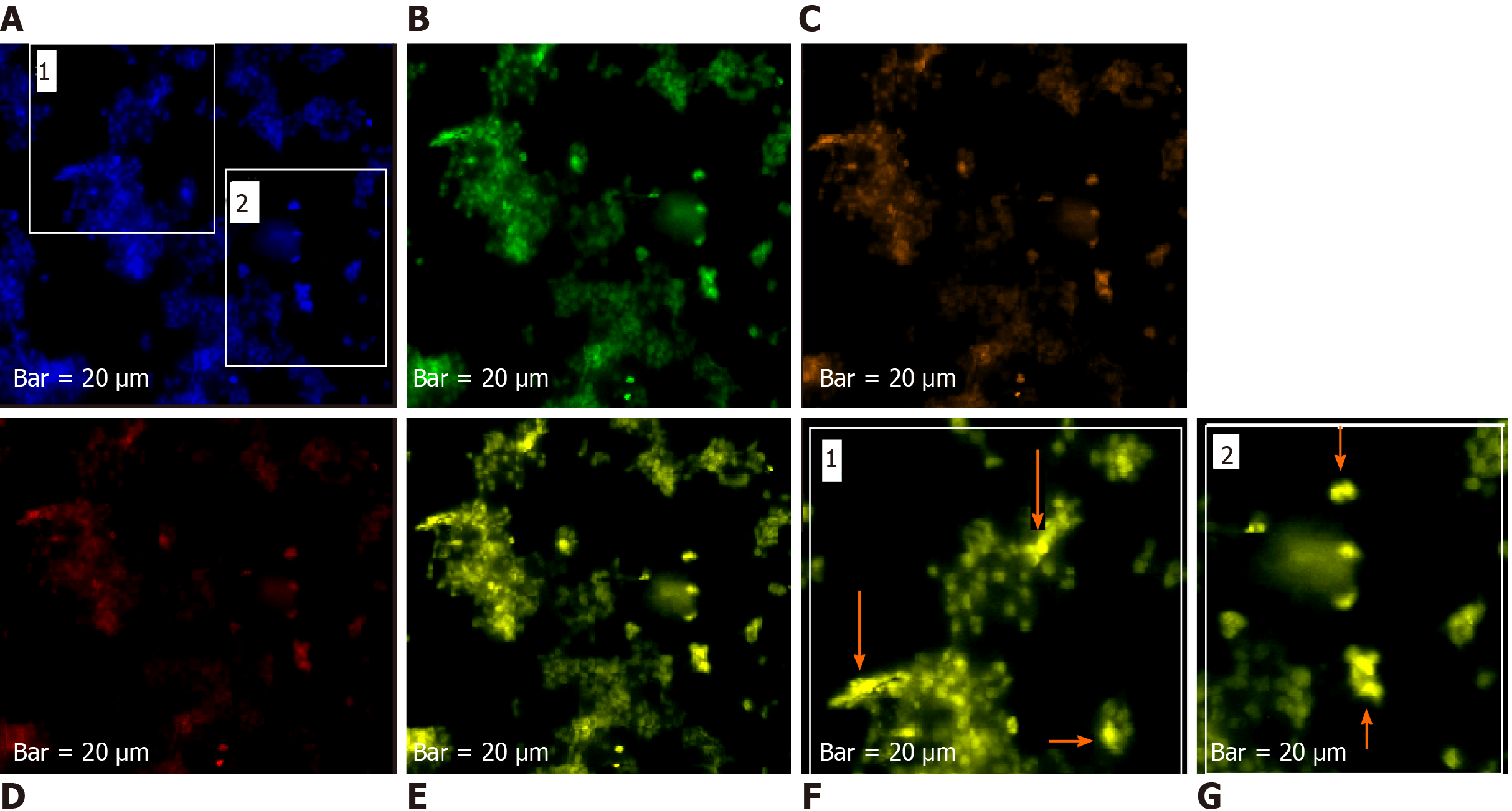
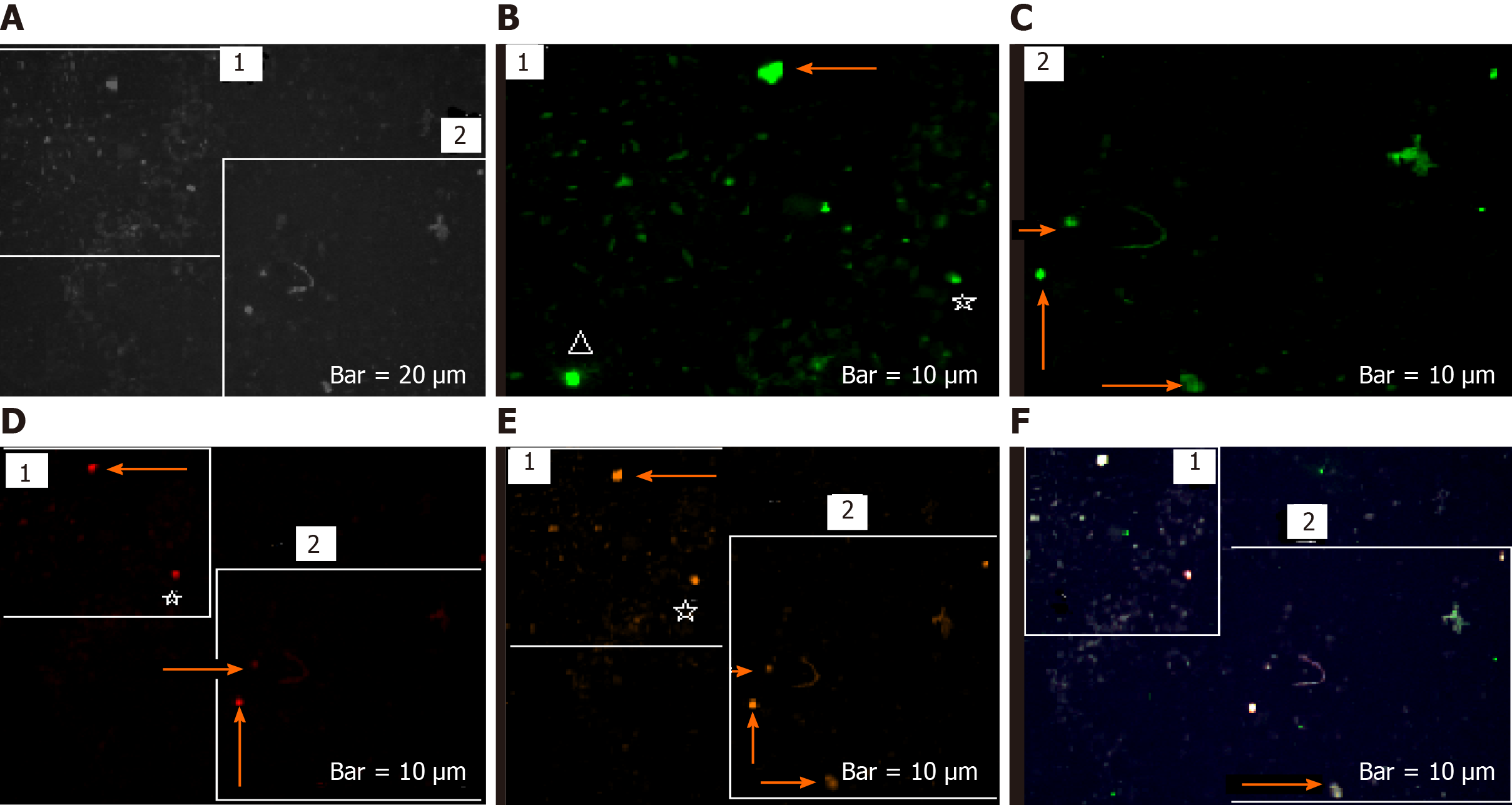
Figure 8 Mode of protein expression and co-expression of C-C chemokine ligand 2, vascular endothelial growth factor, and epidermal growth factor of the circulating tumor cells of patient 13 with meningioma.
C-C chemokine ligand 2 (CCL2), vascular endothelial growth factor (VEGF), and epidermal growth factor (EGF) are conjugated with fluorescein isothiocyanate, R-phycoerythrin, and phycoerythrin–indodicarbocyanine, respectively. A: Leukocytes and circulating tumor cells (CTCs) with 4′,6-diamidino-2-phenylindole; B: Image of the same cells with CCL2; C: Image of the same cells with VEGF; D: Image of the same cells with EGF; E: Co-expression of the CCL2/VEGF/EGF in the same cells; F and G: Co-expression of the CCL2/VEGF/EGF in the selected cropping cells (1 and 2) from A. The arrows are reflective of high protein co-expression within CTCs according the mode of expression in images b-d in the EGF; and the VEGF reflects the lowest expression.
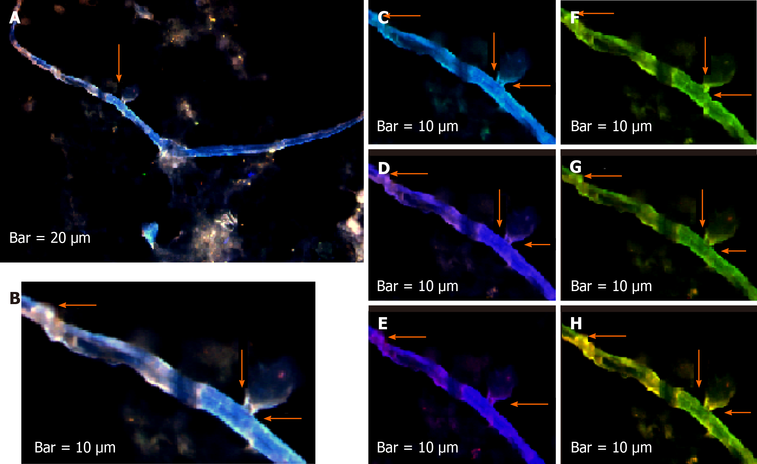
Figure 9 Status of protein expression and co-expression of C-C chemokine ligand 2/vascular endothelial growth factor/epidermal growth factor through migration of circulating tumor cells within tumor territory to the blood stream in a patient with the lung metastatic carcinoma.
C-C chemokine ligand 2 (CCL2), vascular endothelial growth factor (VEGF) and epidermal growth factor (EGF) are conjugated with fluorescein isothiocyanate; R-phycoerythrin, and phycoerythrin–indodicarbocyanine, respectively, in patient 15. A: Merged of 4′,6-diamidino-2-phenylindole (DAPI)/CCL2/EGF/VEGF; B: Cropped section of image a; C: Merged of DAPI/CCL2; D: Merged of DAPI/VEGF; E: Merged of DAPI/EGF; F: Co-expression of CCL2/Veg; G: Co-expression of CCL2/EGF; H: Co-expression of CCL2/VEGF/EGF. Merged of DAPI/CCL2/VEGF/EGF is reflective of co-expression between three proteins; B, F-H: Reflect co-expression of CCL2/VEGF/EGF according to the selected of cropped section from images a (arrow on the top/left). The arrows on the left side of images are referred to the migration of tumor cells from tumor to the blood stream destination through a bubble like channel.
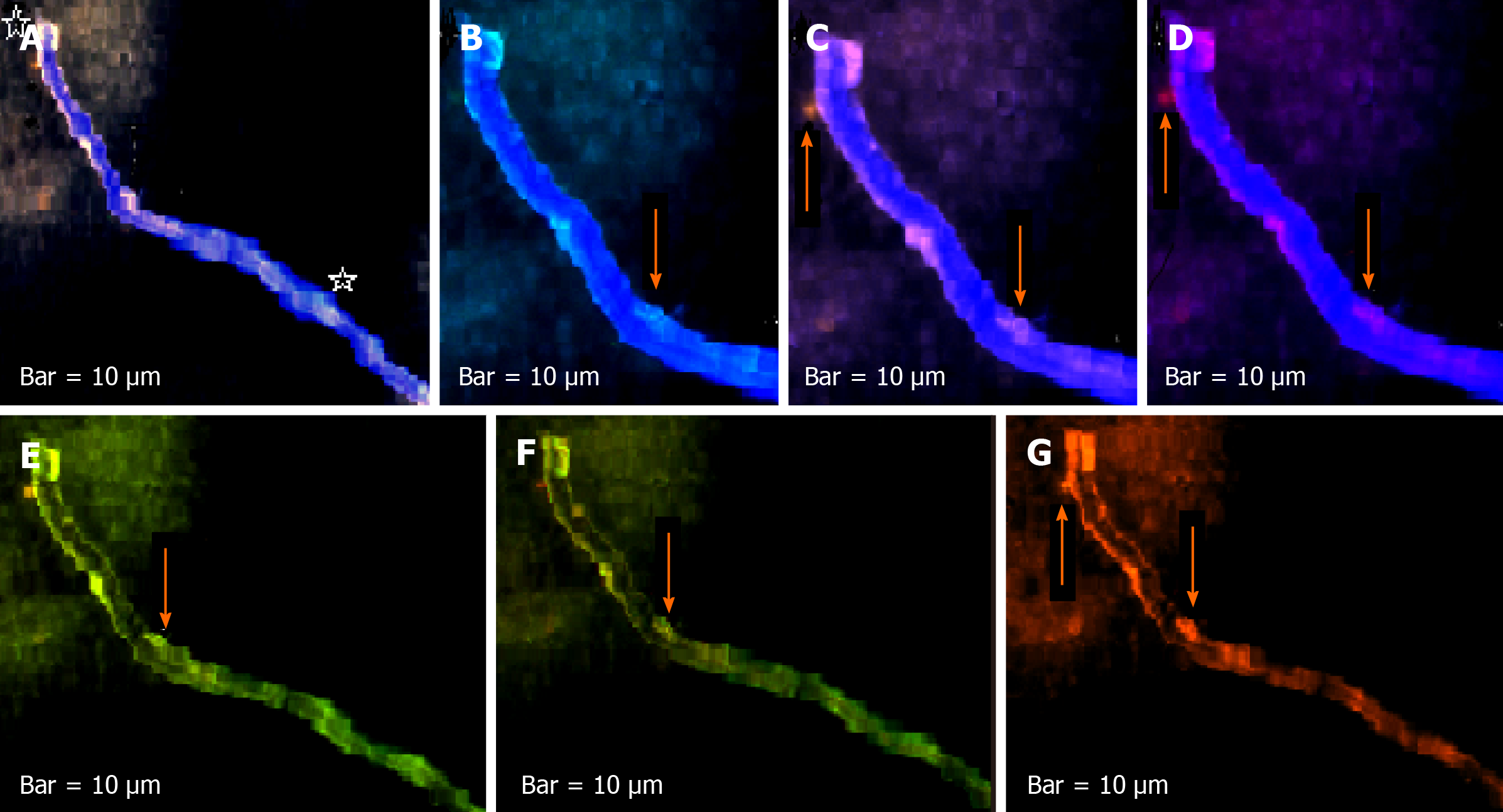
Figure 10 Expression mode of C-C chemokine ligand 2, vascular endothelial growth factor and epidermal growth factor in the migrated vasculature into the blood stream in a patient affected with metastatic brain tumor with breast origin.
C-C chemokine ligand 2 (CCL2), vascular endothelial growth factor (VEGF) and epidermal growth factor (EGF) are conjugated with fluorescein isothiocyanate, R-phycoerythrin, and phycoerythrin–indodicarbocyanine, respectively in patient 17. A: Merged of 4′,6-diamidino-2-phenylindole (DAPI)/CCL2/VEG/EGF; B, C and D: Partially cropped section between two asterisk of image A and present the merged of DAPI/CCL2, DAPI/VEGF and DAPI/EGF respectively; B and C: Indicates the involvement of traceable tumor cells with higher expression than EGF within a migrated vasculature to the blood stream; E, F and G: Present co-expression between CCL2/Veg, CCL2/EGF and EGF/VEGF respectively, there is more harmony between CCL2/Veg and CCL2/Veg.
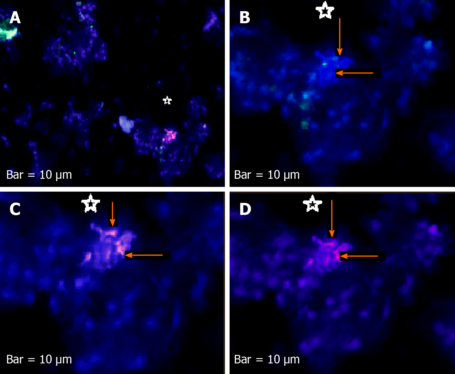
Figure 11 Expression mode of proteins of C-C chemokine ligand 2, vascular endothelial growth factor, and epidermal growth factor in circulating tumor cells from a tumor of patient affected with cerebellar medulloblastoma.
A: Merged image of cells conjugated with 4′,6-diamidino-2-phenylindole (DAPI)/C-C chemokine ligand 2 (CCL2)/vascular endothelial growth factor (VEGF)/epidermal growth factor (EGF) is reflective of remarkable co-expression between VEGF/EGF proteins; B: Merged cells with DAPI/CCL2 indicating the absence of expression in the majority of cells; C: Merged cells with DAPI/VEGF with high expression; D: Merged cells with DAPI/EGF reflect lower expression than VEGF. Asterisks are referred to the cluster of CTCs.
Figure 12 Status of protein expression of neuronal marker, CD133 and vascular endothelial growth factor in the brain circulating tumor cells of patient 1 affected with meningioma.
A: Circulating tumor cells (CTCs) with phase contrast; B and C: Present CTCs with Ne conjugated with fluorescein isothiocyanate; D: Present status of vascular endothelial growth factor (VEGF), conjugated with phycoerythrin–indodicarbocyanine; E: Reflect status of CD133, conjugated with R-phycoerythrin; F: Present the merged image of 4′,6-diamidino-2-phenylindole/Ne/VEGF/CD133, reflective of a remarkable co-expression between protein neuronal markers CD133 and VEGF. Arrows on the top of images B, D1, and E1 reflect more involved cells with high-, very less involved cells with much lower-, and less involved cells with high-expression for Ne, VEGF, and CD133, respectively. The asterisks on the down/right of B, B and E illustrate almost same intensity of expression, but with more CTCs involved in image B for Ne, VEGF, and CD133, respectively. A triangle symbol is reflective of a remarkable high expression for neuronal marker in CTCs. Arrows on the below/left-down of images C2, D2, and E2 present higher expression for Ne than VEGF and CD133. Arrows on the bellow/left-up of images C2, D2, and E2 illustrate low expression for Ne and CD133, and lack of expression for VEGF. The arrows down the images of C2, D2, and E2 illustrate the heterogenic (low and lack) of expression for Ne and CD133 and lack of expression for VEGF.
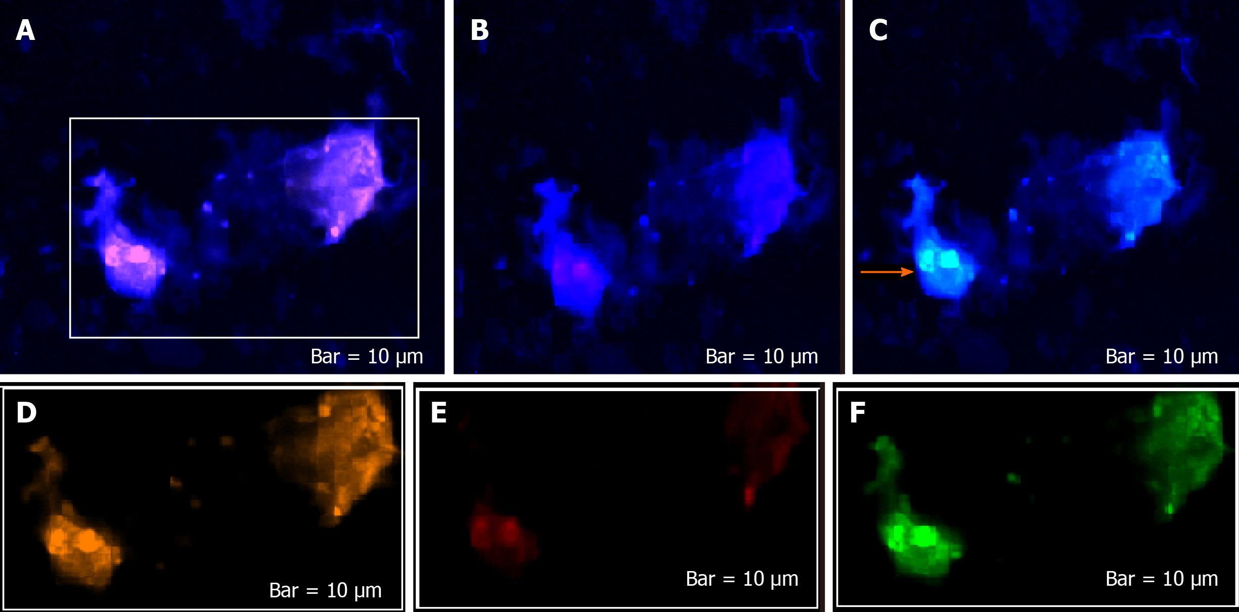
Figure 13 Protein expression of cytokeratin-19, CD45, and cyclin E in circulating tumor cells of a breast-brain metastatic patient.
Circulating tumor cells (CTCs) in patient 17 with: A: DAPI (4′,6-diamidino-2-phenylindole)/cytokeratin-19 (CK-19) [highest/positive protein expression (PE)]; B: DAPI/CD45 (negative PE); C: DAPI/cyclin E (positive PE: Partial at left clone of cells); D, E and F: Conjugated cells with CK-19, CD45, and cyclin E, respectively. Harmonic PE between CK-19 and cyclin E in the clone of CTCs at left side of image in patient 17. Arrows indicate the diverse expression of three proteins.
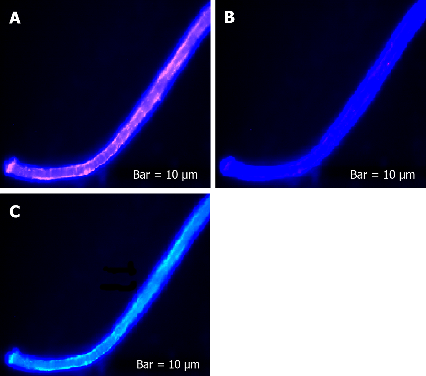
Figure 14 Protein expression of cytokeratin-19, CD45, and cyclin E in circulating tumor cells of a vasculature image of a breast-brain metastatic patient.
Cytokeratin-19 (CK-19), CD45, and cyclin E are conjugated with phycoerythrin-indodicarbocyanine, R-phycoerythrin, and fluorescein isothiocyanate, respectively. A: 4′,6-Diamidino-2-phenylindole (DAPI)/CK-19 [highest/positive protein expression (PE)]; B: DAPI/CD45 (negative PE); C: DAPI/cyclin E (positive PE: Partial at left clone of cells). Harmonic PE between CK-19 and cyclin E, arrows indicate lack of cyclin E expression in the middle of vasculature in patient 17.
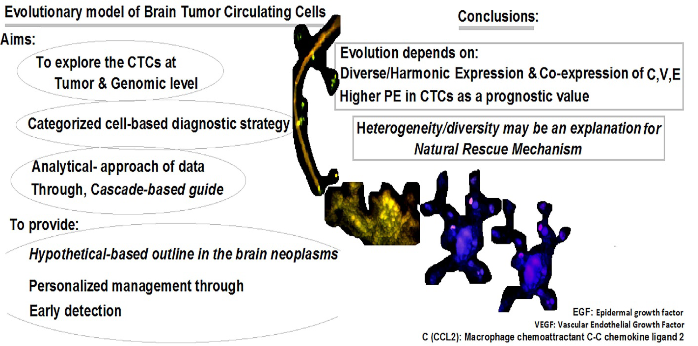
Figure 15 Manuscript at a glance: Evolutionary model of brain tumor circulating cells.
CTCs: Circulating tumor cells; PE: Protein expression.
- Citation: Mehdipour P, Javan F, Jouibari MF, Khaleghi M, Mehrazin M. Evolutionary model of brain tumor circulating cells: Cellular galaxy. World J Clin Oncol 2021; 12(1): 13-30
- URL: https://www.wjgnet.com/2218-4333/full/v12/i1/13.htm
- DOI: https://dx.doi.org/10.5306/wjco.v12.i1.13


