Copyright
©The Author(s) 2019.
World J Clin Oncol. Apr 24, 2019; 10(4): 192-200
Published online Apr 24, 2019. doi: 10.5306/wjco.v10.i4.192
Published online Apr 24, 2019. doi: 10.5306/wjco.v10.i4.192
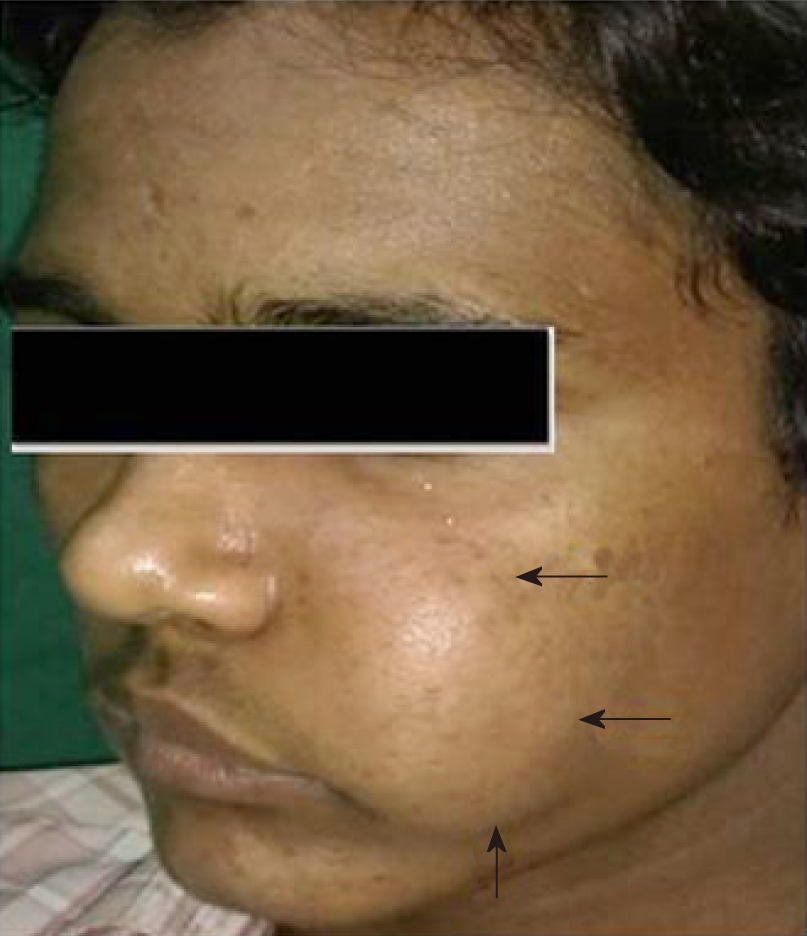
Figure 1 Clinical image shows a diffuse swelling over left side of the face causing facial asymmetry.
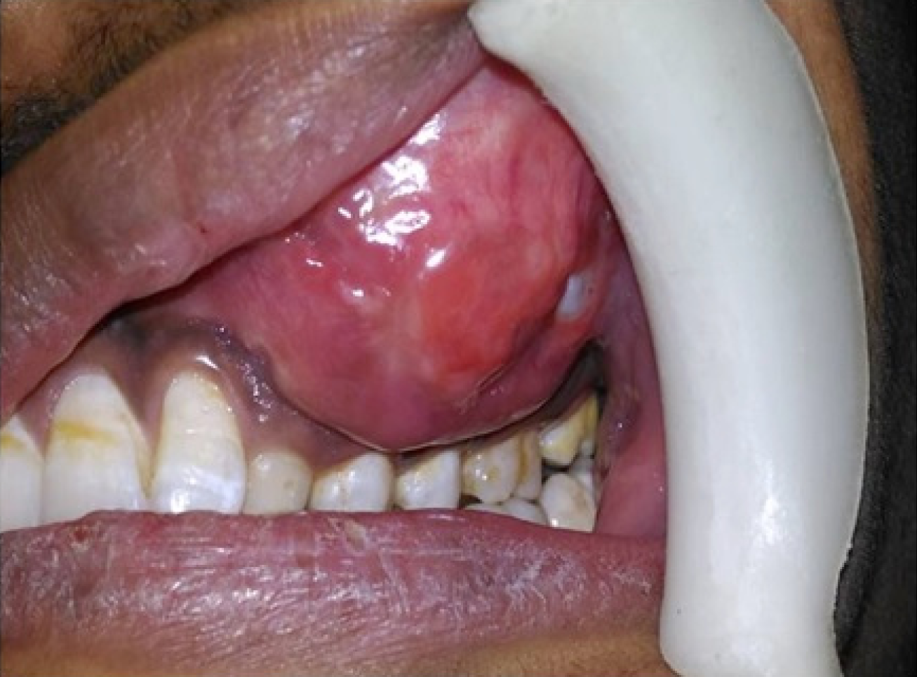
Figure 2 Intraoral view shows a swelling of approximately 7 cm × 5 cm in size over maxillary left region obliterating the buccal vestibule.
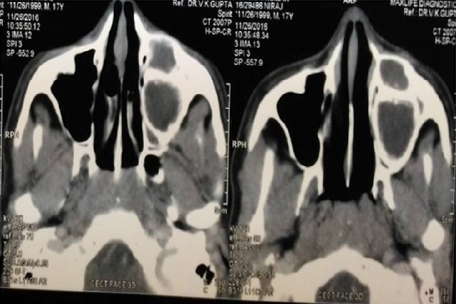
Figure 3 Computed tomography scan shows mixed radiopaque radiolucent lesion over left maxillary region.
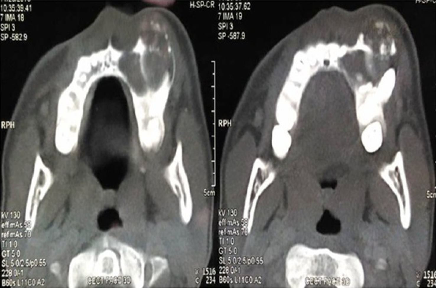
Figure 4 Computed tomography scan shows expansion, thinning and perforation of buccal cortical plate with multiple intermittent radiopaque flecks within the radiolucent area.
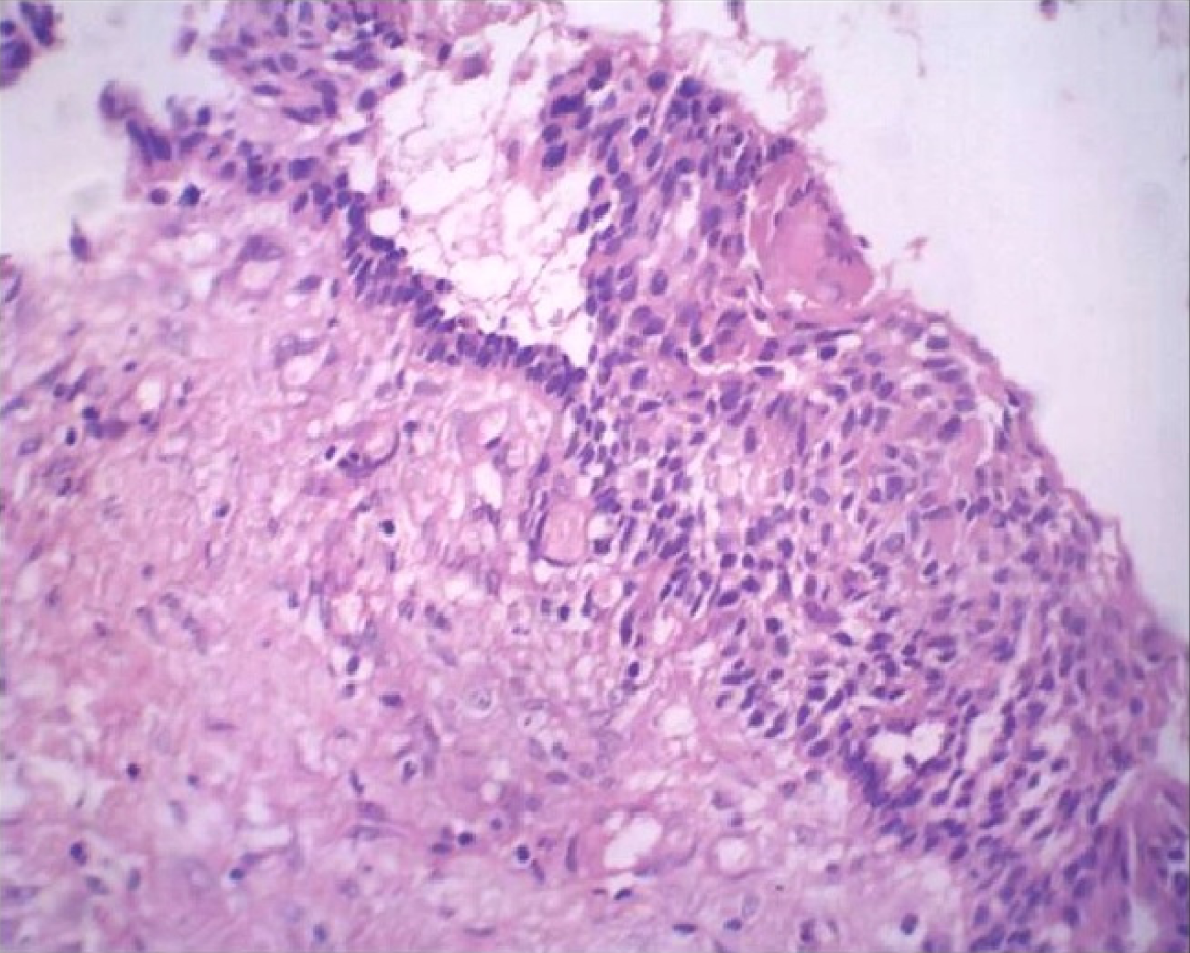
Figure 5 Histopathological image shows odontogenic epithelium with tall columnar basal cell layer, stellate reticulum like cells and ghost cells (H and E, ×100).
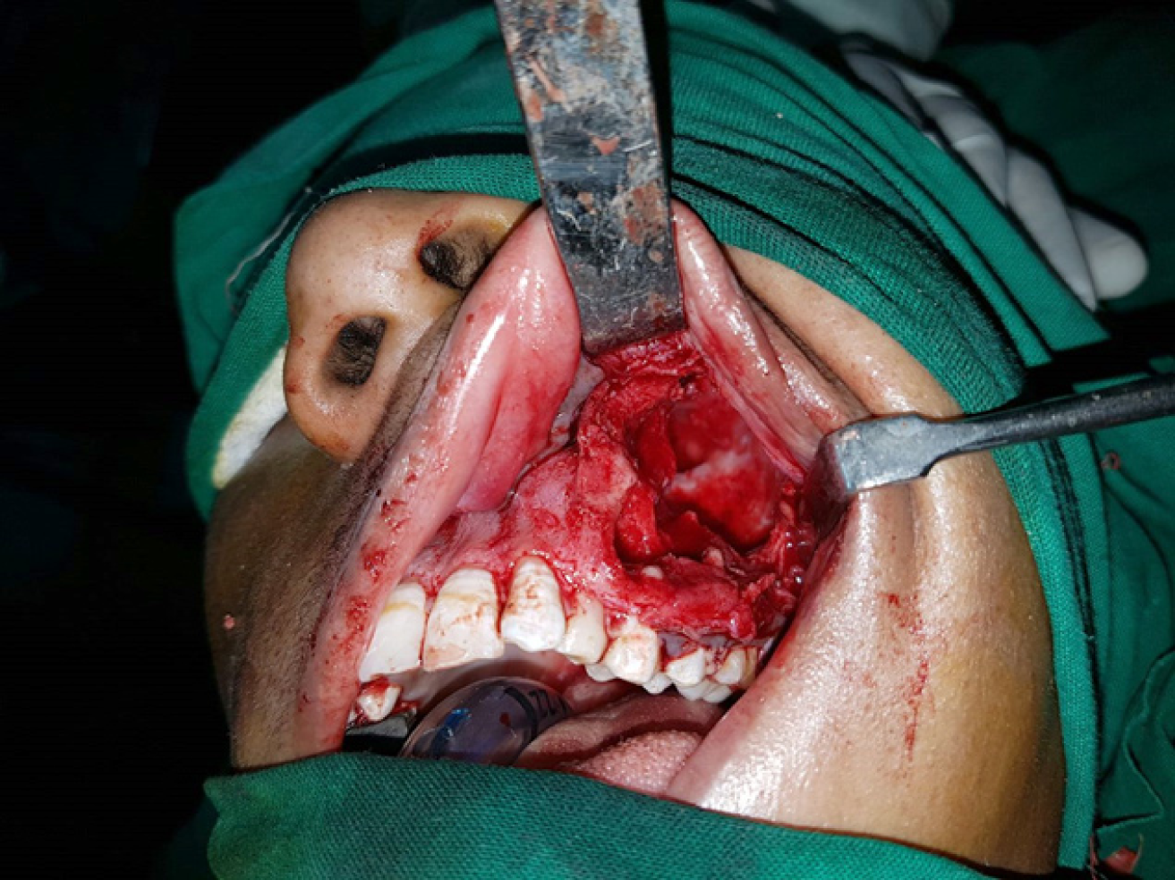
Figure 6 Clinical image of the intraoperative site showing segmental resection being carried out.
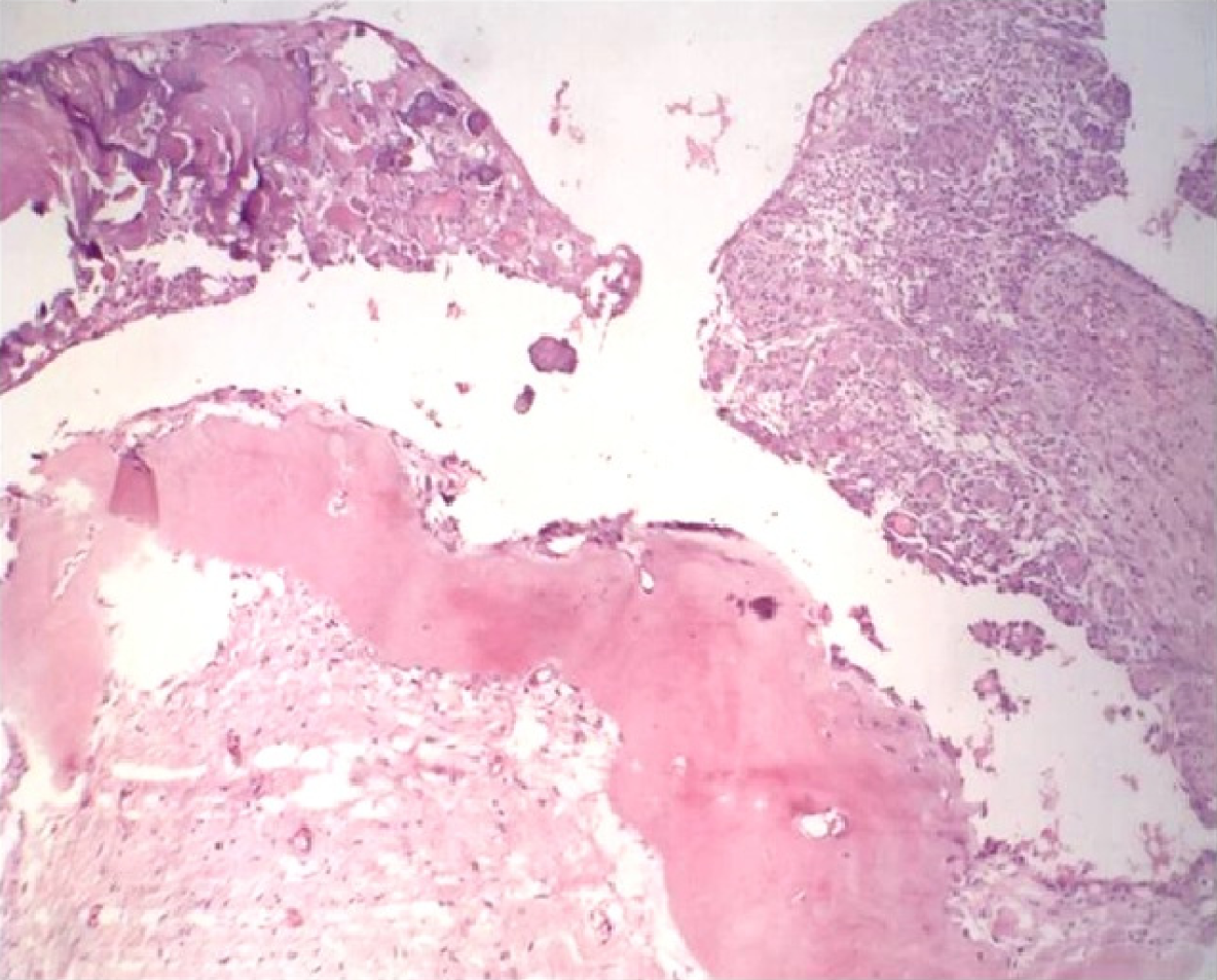
Figure 7 Histopathological image shows aggregates of eosinophilic ghost cells with irregular calcifications in the epithelium along with large areas of dentinoid seen in the subjacent connective tissue (H and E, ×40).
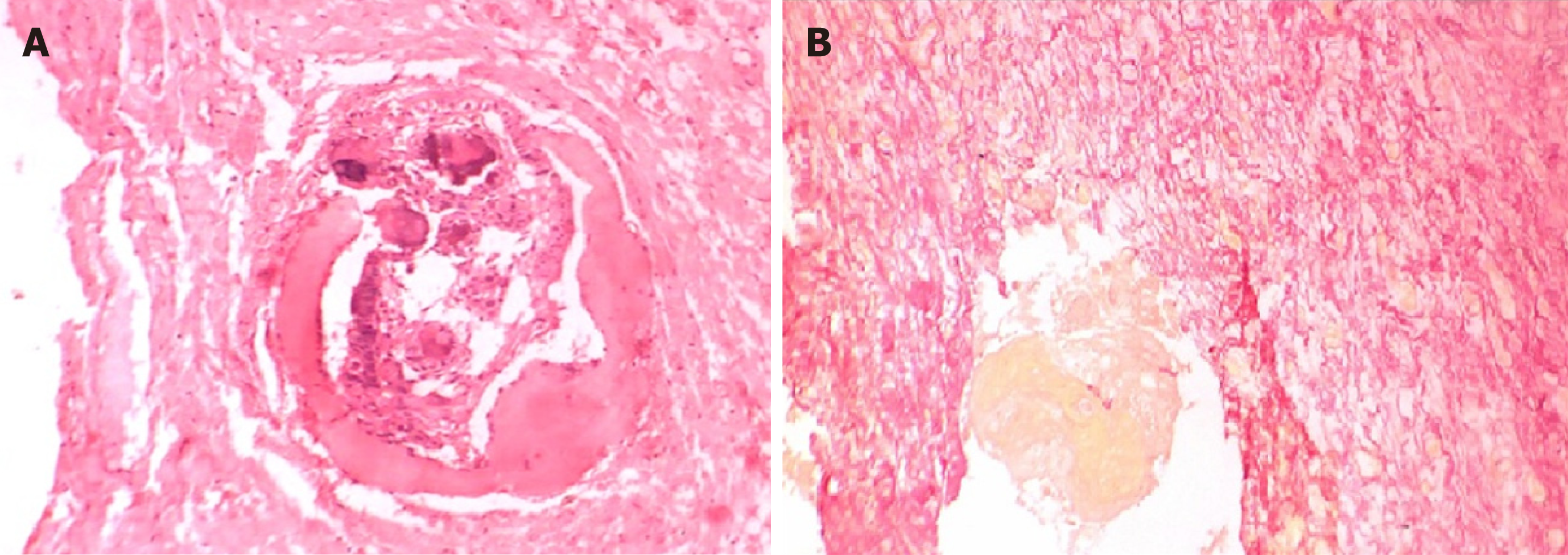
Figure 8 Histopathological image.
A: It shows dystrophic calcification in epithelium and pinkish areas demonstrating dentinoid seen on special staining (van Gieson, ×100); B: Yellow area showing aggregates of ghost cells (van Gieson, ×100).
- Citation: Patankar SR, Khetan P, Choudhari SK, Suryavanshi H. Dentinogenic ghost cell tumor: A case report. World J Clin Oncol 2019; 10(4): 192-200
- URL: https://www.wjgnet.com/2218-4333/full/v10/i4/192.htm
- DOI: https://dx.doi.org/10.5306/wjco.v10.i4.192


