Copyright
©The Author(s) 2019.
World J Clin Oncol. Dec 24, 2019; 10(12): 402-408
Published online Dec 24, 2019. doi: 10.5306/wjco.v10.i12.402
Published online Dec 24, 2019. doi: 10.5306/wjco.v10.i12.402
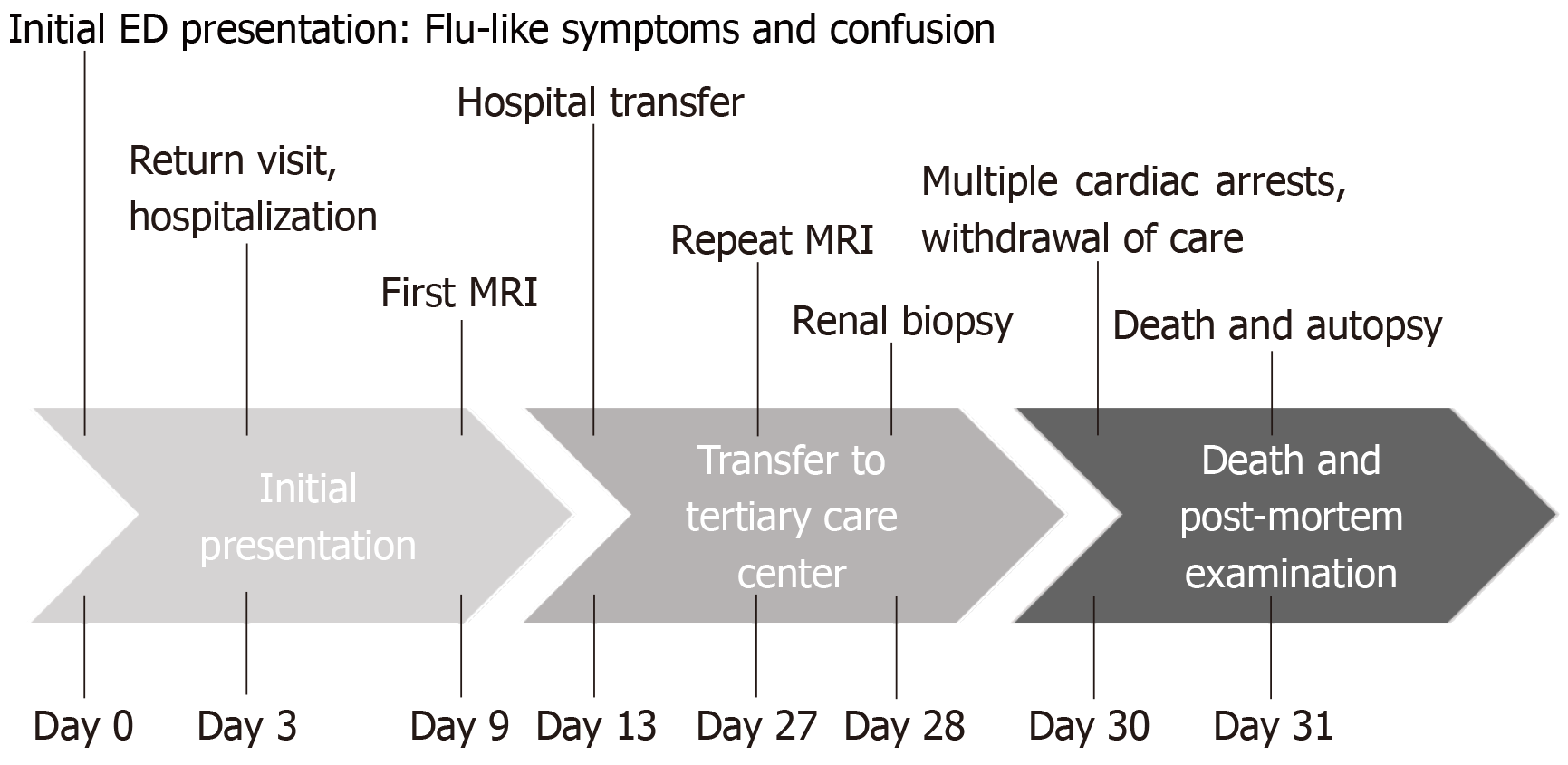
Figure 1 Case timeline.
MRI: Magnetic resonance imaging; ED: Emergency department.
Figure 2 Magnetic resonance imaging brain findings.
Evolution of central nervous system changes highlighting the corpus callosum involvement and parenchymal abnormalities related to intravascular large B-cell lymphoma. A: Normal magnetic resonance imaging (MRI) taken August 21, 2017; B: MRI brain 3 m later at presentation revealing splenium enhancement also known as the “boomerang sign[8]”; C: MRI taken 18 d following the MRI shown in B with continued evolution of splenium enhancement as well as progressive parenchymal enhancement posteriorly.
Figure 3 Histopathologic analysis.
A: Post-mortem renal parenchyma high power view demonstrating lymphovascular cells distending the lumen of a vessel lined by epithelial cells (original magnification 40×); B: Immunohistochemical stain from an ante-mortem renal biopsy for CD20+ highlighting lymphocytes restricted to vascular spaces (original magnification 20×); C: Post-mortem pituitary section revealing extensive lymphocyte involvement of lymphovascular spaces (original magnification 20×); D: Intravascular large B-cell lymphoma within cerebral vessels with focal sludging (original magnification 20×).
- Citation: D’Angelo CR, Ku K, Gulliver J, Chang J. Intravascular large B-cell lymphoma presenting with altered mental status: A case report. World J Clin Oncol 2019; 10(12): 402-408
- URL: https://www.wjgnet.com/2218-4333/full/v10/i12/402.htm
- DOI: https://dx.doi.org/10.5306/wjco.v10.i12.402


