Copyright
©The Author(s) 2015.
World J Gastrointest Pharmacol Ther. Aug 6, 2015; 6(3): 89-95
Published online Aug 6, 2015. doi: 10.4292/wjgpt.v6.i3.89
Published online Aug 6, 2015. doi: 10.4292/wjgpt.v6.i3.89
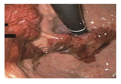
Figure 1 Endoscopic image of the gastric cardia demonstrating Kaposi’s sarcoma lesion extension from the gastric body.
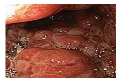
Figure 2 Endoscopic image of the gastric body showing an extensive, infiltrative, and circumferential Kaposi’s sarcoma mass involving the entire body.
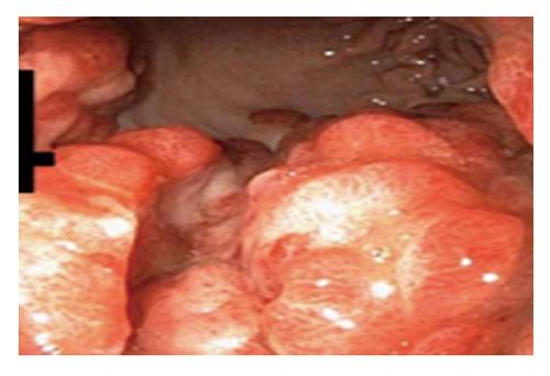
Figure 3 Endoscopic image of the gastric antrum with Kaposi’s sarcoma lesion infiltration from the body.
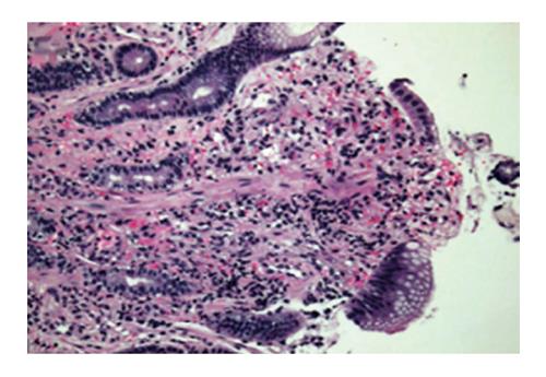
Figure 4 Hematoxylin and eosin stain of slit-like spaces with spindle cell proliferation and associated red blood cell extravasation seen in the lamina propria.
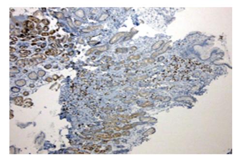
Figure 5 Immunohistochemical stain for human herpes virus 8 showing a strongly positive latent nuclear antigen staining of the spindle cells along the slit-like spaces.
- Citation: Lee AJ, Brenner L, Mourad B, Monteiro C, Vega KJ, Munoz JC. Gastrointestinal Kaposi’s sarcoma: Case report and review of the literature. World J Gastrointest Pharmacol Ther 2015; 6(3): 89-95
- URL: https://www.wjgnet.com/2150-5349/full/v6/i3/89.htm
- DOI: https://dx.doi.org/10.4292/wjgpt.v6.i3.89


