Copyright
©The Author(s) 2018.
World J Gastrointest Pathophysiol. Feb 15, 2018; 9(1): 1-7
Published online Feb 15, 2018. doi: 10.4291/wjgp.v9.i1.1
Published online Feb 15, 2018. doi: 10.4291/wjgp.v9.i1.1
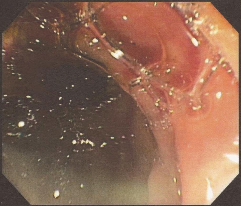
Figure 1 Pus seen extruding from the ampulla of Vater.
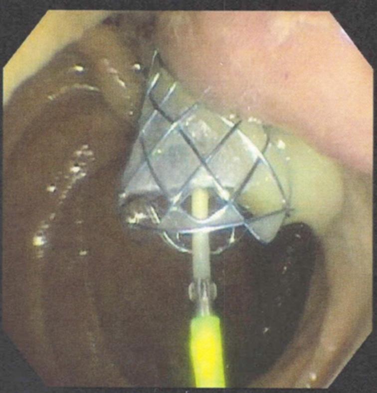
Figure 2 Drainage of pus after biliary stenting during endoscopic retrograde cholangiopancreatography.
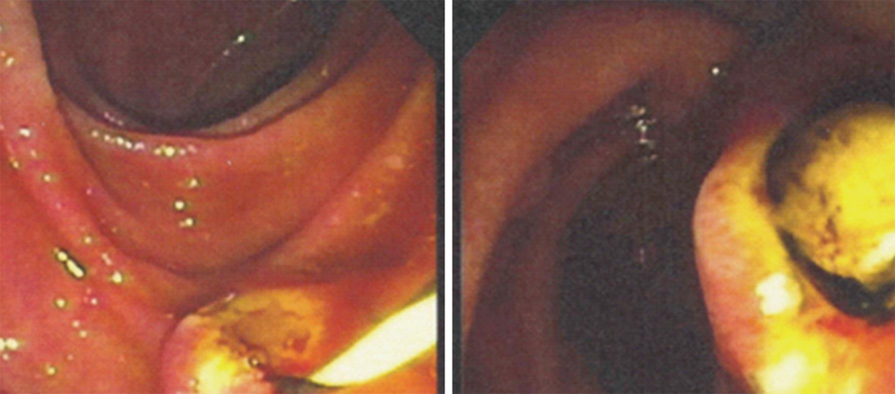
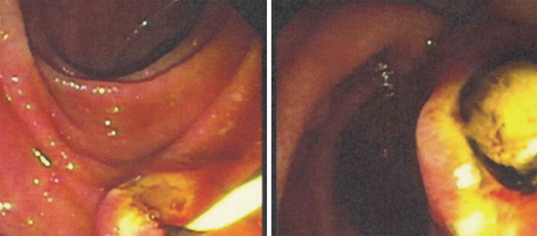
Figure 3 Biliary sphincterotomy followed by stone extraction.
Figure 4 Biliary stone extraction followed by stent placement.
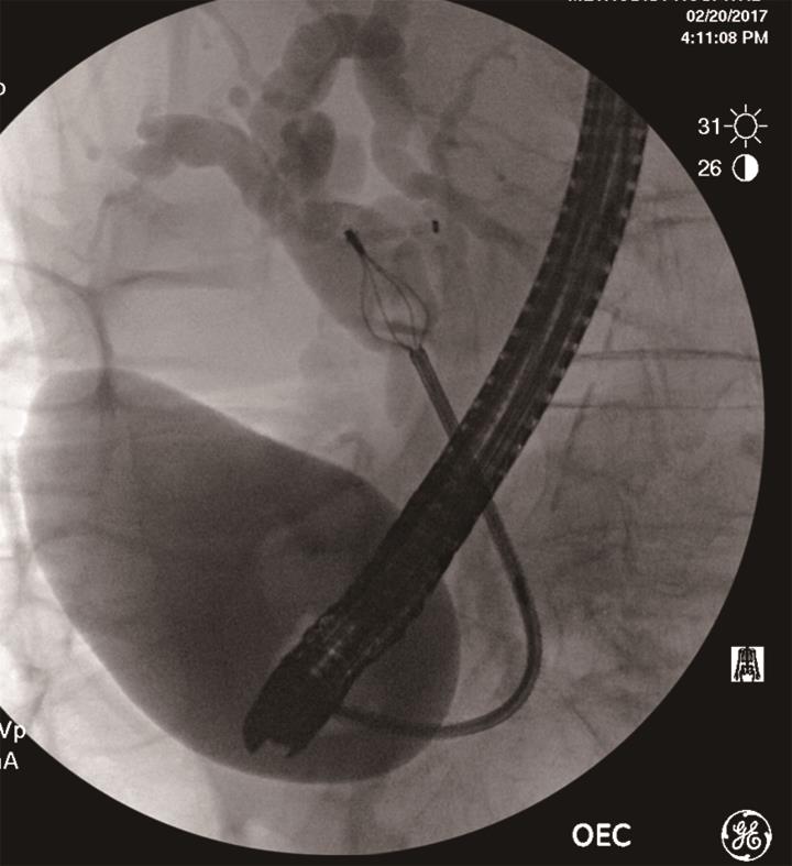
Figure 5 Fluoroscopy showing lithotripsy basket-assisted stone extraction.
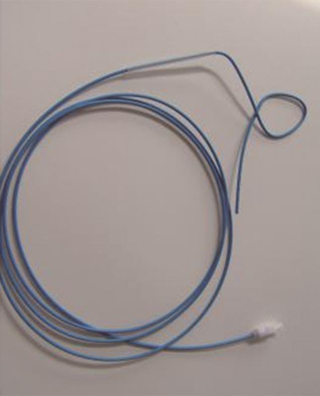
Figure 6 Nasobiliary catheter.
- Citation: Ahmed M. Acute cholangitis - an update. World J Gastrointest Pathophysiol 2018; 9(1): 1-7
- URL: https://www.wjgnet.com/2150-5330/full/v9/i1/1.htm
- DOI: https://dx.doi.org/10.4291/wjgp.v9.i1.1


