Published online Feb 28, 2023. doi: 10.4329/wjr.v15.i2.32
Peer-review started: May 29, 2022
First decision: August 22, 2022
Revised: September 23, 2022
Accepted: February 13, 2023
Article in press: February 13, 2023
Published online: February 28, 2023
Processing time: 270 Days and 6.3 Hours
Coronary computed tomography angiography (CCTA) is the preferred non-invasive examination method for coronary heart disease. However, the radiation from computed tomography has become a concern since public awareness of radiation hazards continue to increase.
To explore the value of multiple dose reduction techniques for CCTA.
Consecutive normal and overweight patients were prospectively divided into two groups: Group A1, patients who received multiple dose reduction scans (n = 82); and group A2, patients who received conventional scans (n = 39). The scan parameters for group A1 were as follows: Isocentric scan, tube voltage = 80 kV, and tube current control using 80% smart milliampere. The scan parameters for group A2 were as follows: Normal position, tube voltage = 100 kV, and smart milliampere.
The average effective doses (EDs) for groups A1 and A2 were 1.13 ± 0.35 and 3.36 ± 1.30 mSv, respectively. There was a statistically significant difference in ED between the two groups (P < 0.01). Furthermore, noise was significantly lower, and both signal-to-noise ratio and contrast signal-to-noise ratio were higher in group A2 when compared to group A1 (P < 0.01). Moreover, the subjective image quality (IQ) scores were excellent in both groups, in which there was no signi
Multiple dose reduction scan techniques can significantly decrease the ED of patients receiving CCTA examinations for clinical diagnosis.
Core Tip: Coronary computed tomography angiography (CCTA) is the preferred non-invasive examination method for coronary heart disease (CHD). The present study was the first to combine multiple dose reduction techniques, including narrow acquisition window, low tube voltage, lower tube current, and isocentric scanning, to decrease CCTA radiation exposure of patients with suspected CHD. The radiation dose for the group with multiple dose reduction was approximately 33.63% (1.13 ± 0.35/3.36 ± 1.30) of the dose associated with the conventional method.
- Citation: Hu XL, Huang PK, Zhang M, Chen J, Xiao MQ. Effects of combining multiple dose reduction techniques on coronary computed tomography angiography. World J Radiol 2023; 15(2): 32-41
- URL: https://www.wjgnet.com/1949-8470/full/v15/i2/32.htm
- DOI: https://dx.doi.org/10.4329/wjr.v15.i2.32
Coronary computed tomography angiography (CCTA) is the preferable non-invasive method to check for coronary heart disease (CHD)[1,2]. In terms of technology, CCTA has advanced significantly during the last 20 years. For those who suspect they may have CHD, CCTA has largely taken the role of conventional invasive coronary angiography since it is so effective at detecting moderate to severe coronary artery stenosis[2]. But as individuals become increasingly conscious of the risks associated with radiation, computed tomography (CT) radiation has risen to the forefront of discussion[3].
Numerous dose-reduction techniques have been developed as a result of the advancement of computer technology, including prospective electrocardiography gating, iterative reconstruction, personalized scanning, as well as ”smart milliampere” (the CT can automatically calculate the tube current based on the position scan) have been developed[3,4]. Furthermore, with the emergence of high-end CT machines, adjusting the tube voltage and tube current has become a useful approach for dose reduction in CCTA scanning[1,5,6]. Some organizations have reported the application of dual-source CT (Siemens Healthcare, Malvern, PA, United States) and GE gem energy spectrum CT (Medical Systems, Milwaukee, WI, United States) to reduce the tube voltage to 80 kV and manage the tube current with smart milliamperes, thereby decreasing the effective dose (ED) to a significant degree[7,8]. Furthermore, previous researchers have employed single dose reduction techniques to minimize the CCTA radiation dose. However, there are limitations in reducing the overall ED. Therefore, more advanced methods are warranted to further decrease the radiation exposure during CCTA.
Isocentric scanning has been proven as an effective approach to reduce radiation exposure in patients receiving CCTA examinations[9,10]. The direction of the beam in the CT is fixed in the middle of the CT frame. The beam in multislice spiral CT has a cone-like form. More light would reach the system through a spherical tube and frame-hole-center-scanning detector, which would enhance the quality of the objective picture [image quality (IQ)]. When an object is being scanned isocentrically, it aligns with the gantry’s empty center. This technology has been heavily utilized by all varieties of CT. Additionally, it has been shown that using this technique in CT may lower the ED of scans for organs that are not in the middle[11,12].
To our knowledge, the present study was the first to combine multiple dose reduction techniques, including narrow acquisition window, low tube voltage, lower tube current, and isocentric scanning, in order to decrease the CCTA radiation exposure of patients with suspected CHD. The present study aimed to determine whether multiple dose reduction approaches can efficiently reduce the radiation exposure while maintaining the IQ in CCTA examinations.
The present prospective research was approved by the institutional review board of Guangdong Provincial Hospital of Traditional Chinese Medicine Medical Ethics Committee (BF2020-229-01), and all patients provided written informed consent. Between November 2020 and August 2021, consecutive patients with clinically suspected CHD were screened for inclusion in the present study. The inclusion criteria were as follows: Age ≥ 18-years-old; heart rate < 70 bpm; and body mass index (BMI): 18.0-29.9 kg/m2. Patients with pacemakers, severe respiratory artifacts, metal implants within the scan range, allergies to contrast media or Betaloc, or a history of cardiac tumors or cardiac surgery were excluded.
The study population comprised 118 patients with CHD. These patients were classified according to their BMI, as described above, and divided into two groups [the groups were classified as normal or overweight (BMI: 18.0-29.9 kg/m2)[13], BMI = weight (kg)/height (m)2]: Multi-modality normal group (group A1, n = 82) and conventional normal group (group A2, n = 39). Coronary artery segmentation was performed according to the 15-segment coronary artery segmentation method developed by the American Heart Association[1].
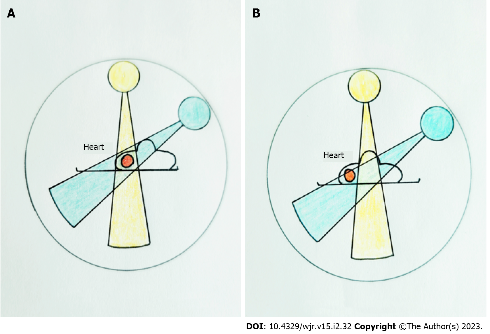
The CCTA examination was performed using the Canon 320-row dynamic CT system (Aquilion ONE Genesis, Canon Medical Systems). Prospective electrocardiography gating was used to reduce the radiation dose[6]. The scanning range was extended 140 mm upwards, starting from the lower edge of the heart. For group A1, the isocentric scan was performed with the patient supine and their body shifted to the right (Figure 1). Before the CCTA scan, an ultrasound examination was performed to mark the leftmost and rightmost edges of the heart on the body surface, and perpendicular lines were placed along the leftmost and rightmost edges of the heart. The centerline of the two vertical lines was used as the vertical positioning line, and the horizontal axillary centerline was used as the horizontal positioning line.
Patients in group A2 were scanned using the conventional method, with the patient placed in the natural supine position. Similarly, the horizontal axillary centerline was used for the horizontal positioning, and the median line was used as the vertical positioning line. The scan parameters were as follows: Group A1, tube voltage = 80 kV, 80% tube current, smart milliampere setting; group A2, tube voltage = 100 kV, tube current control using milliampere (Table 1).
| Dose group | Conventional group | Multi-modality group |
| Tube voltage, kV | 100 | 80 |
| Tube current, mA | Smart milliampere | 80% smart milliampere |
| D-FOV, mm | 220 | 220 |
| Rotation time, s | 0.275 | 0.275 |
| Thickness, mm | 0.5 | 0.5 |
| Interval, mm | 0.5 | 0.5 |
| AIDR3D | Standard EU10 | Standard EU10 |
| Scanning method | ECG gating | ECG gating |
| Cardiac cycle | 30%-80% | 60%-80% |
| Scan position | Conventional | Isocentric |
| Scan length | 140 mm | 140 mm |
All patients provided a written informed consent before participating in the study. Heart rate and blood pressure were measured at rest before the examination. Patients with a heart rate of > 70 bpm were given metoprolol (Betaloc, 25-100 mg). These patients were randomly assigned into two groups (groups A1 and A2) at a ratio of 2:1. Before the examination, these patients were administered with 0.5 mg nitroglycerine tablets.
The CT dose index volume (mGy) and dose length product (mGy × cm) were automatically calculated by the scanner software for all CT protocols. Then, the dose length product was multiplied by a conversion coefficient (k) to determine the ED (ED = dose length product × k, k = 0.014)[13] for each patient (Table 2).
| Group, n | Sex, F/M | Age, yr | BMI, kg/m2 | CTDvol, mGy | DLP, mGy × cm | ED, mSv |
| A1, n = 82 | 31/51 | 59.59 ± 9.66 | 23.06 ± 1.95 | 5.89 ± 1.85 | 80.58 ± 24.80 | 1.13 ± 0.36 |
| A2, n = 36 | 19/20 | 64.15 ± 13.28 | 22.58 ± 1.96 | 17.92 ± 8.02 | 250.90 ± 112.25 | 3.36 ± 1.30 |
| P value | 0.43 | 0.17 | 0.89 | 0.00 | 0.00 | 0.00 |
All images were reconstructed using Adaptive Iterative Dose Reduction Using Three Dimensional Processing. The scanning parameters were as follows: Scan length, 140 mm; slice thickness, 0.5 mm; reconstruction field of view, 220 mm; kernel, EU10 (Table 2).
The objective IQ was evaluated by two experienced cardiovascular radiologists (MQ Xiao, 17 years of experience; PK Huang, 13 years of experience), who were blinded to the scan and reconstruction parameters. A circular region of interest (ROI), with a diameter of 1 cm, was placed within the cortical part of the main bronchus. The coronary artery attenuation values for the proximal ROIs of the left main coronary artery and right coronary artery were measured. The ROI was selected as large as possible, and the vascular wall, vascular calcifications, and non-calcified plaques and artifacts were excluded. The average coronary artery attenuation was equal to the average value of the left main coronary artery and proximal right coronary artery. The ROI measurement was performed on the adjacent myocardial fat. This was repeated three times at each location, and averaged to ensure data consistency. The CT value standard deviation for the main bronchus, and average attenuation values for the coronary artery and pericardial fat were calculated by averaging the values obtained by the two observers. Noise was calculated as the standard deviation of the CT value of the main bronchus[14]. The signal-to-noise ratio and contrast signal-to-noise ratio (CNR) were calculated as follows: Signal-to-noise ratio = average main bronchus CT value/image noise; CNR = (average attenuation of coronary artery - perivascular fat attenuation)/image noise[15].
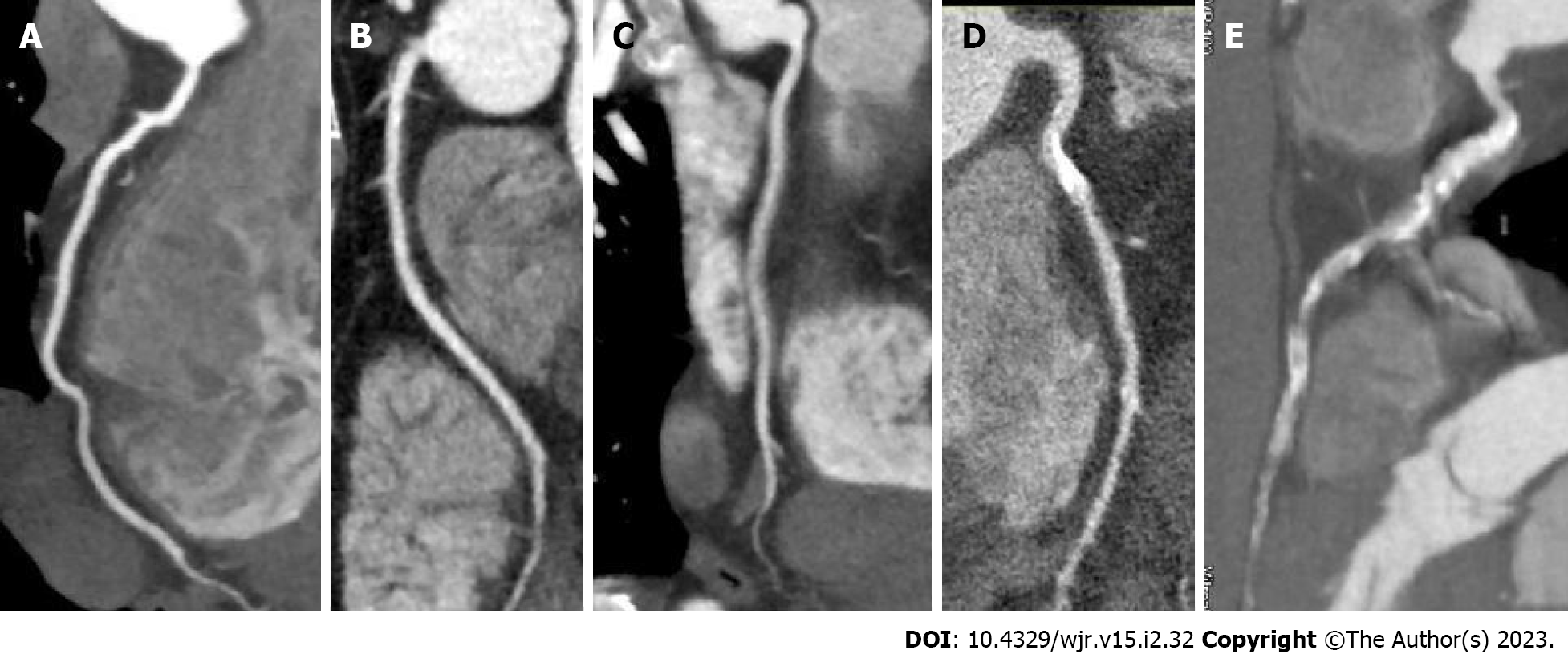
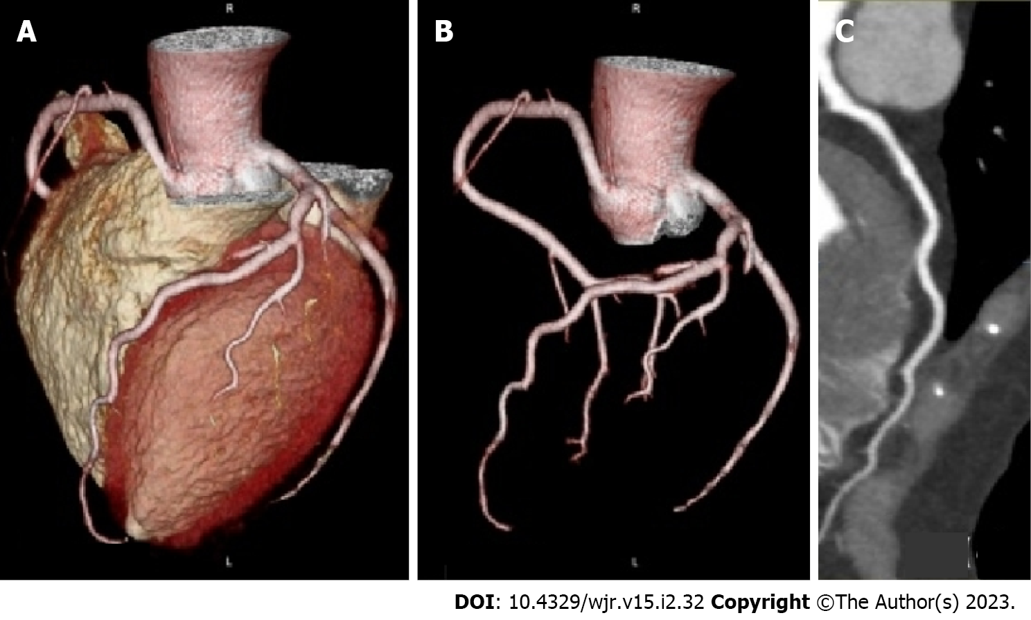
Coronary artery segmentation was performed according to the 15-segment coronary artery segmentation method developed by the American Heart Association[1]. Two radiologists with 18 years (MQ Xiao) and 13 years (PK Huang) of experience in cardiovascular medicine conducted independent evaluations for coronary arteries with a diameter of ≥ 1.5 mm. For inter-rater disagreements, a consensus was reached through consultation. Furthermore, a 5-point Likert scale was used[13] as follows: 5 = excellent (sharp, smooth contours of the vascular wall and no streaking or radiating artifacts); 4 = good (slight irregularities on the contours and few streaks or radiating artifacts); 3 = fair (blurred and irregular contour of the vascular wall and numerous streaks or radiating artifacts); 2 = poor (deformation of the vascular wall and various artifacts); 1 = very poor (obvious deformation of the vascular wall and extensive artifacts). Images with IQ scores of 3-5 satisfied the requirements for the diagnostic assessment (Figure 2).
The study population comprised 118 patients (71 male and 47 female patients), who were within 37-87-years-old (mean ± SD: 61.06 ± 9.10 years).
These normal and overweight patients were divided into two groups: Group A1, patients who underwent multiple dose reduction scan techniques (n = 82); and group A2, patients who underwent the conventional scan technique (n = 39). Sex, age, and BMI did not significantly vary across the groups (P is between 0.06 and 0.43; Table 2).
The average ED of group A1 was 1.13 ± 0.35 mSv, whereas the average ED of group A2 was 3.36 ± 1.30 mSv. The difference between the ED of these two groups was statistically significant (all P less than 0.01). Additionally, the average noise in group A2 was much lower than group A1 (all P more than 0.05; Table 3), and the signal-to-noise ratio and CNR in group A2 were significantly higher than group A1. Additionally, group A1 had considerably higher CT values than group A2 for the right coronary artery root, left main coronary artery, and ascending aortic root (P less than 0.05).
| Group, n | Aortic root, HU | Noise | ||||
| A1, n = 82 | 512.18 ± 108.22 | 477.20 ± 117.93 | 485.88 ± 4100.40 | 15.52 ± 4.73 | 66.00 ± 25.30 | 41.32 ± 16.13 |
| A2, n = 39 | 601.92 ± 125.34 | 534.67 ± 117.33 | 546.99 ± 109.30 | 19.29 ± 6.26 | 54.28 ± 17.72 | 38.45 ± 12.06 |
| P value | 0.07 | 0.32 | 0.37 | 0.00 | 0.00 | 0.00 |
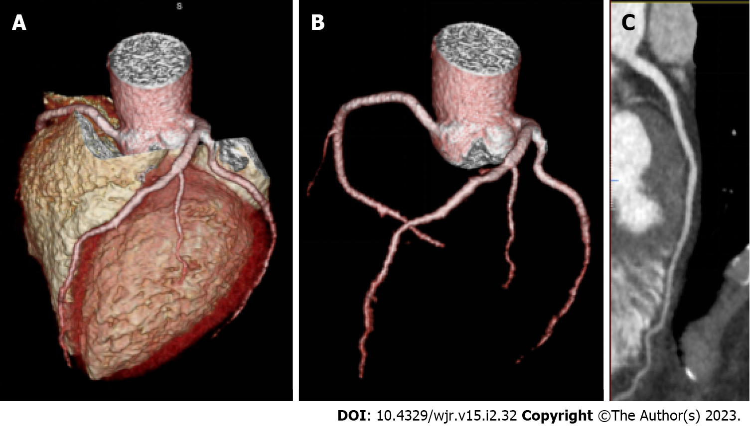
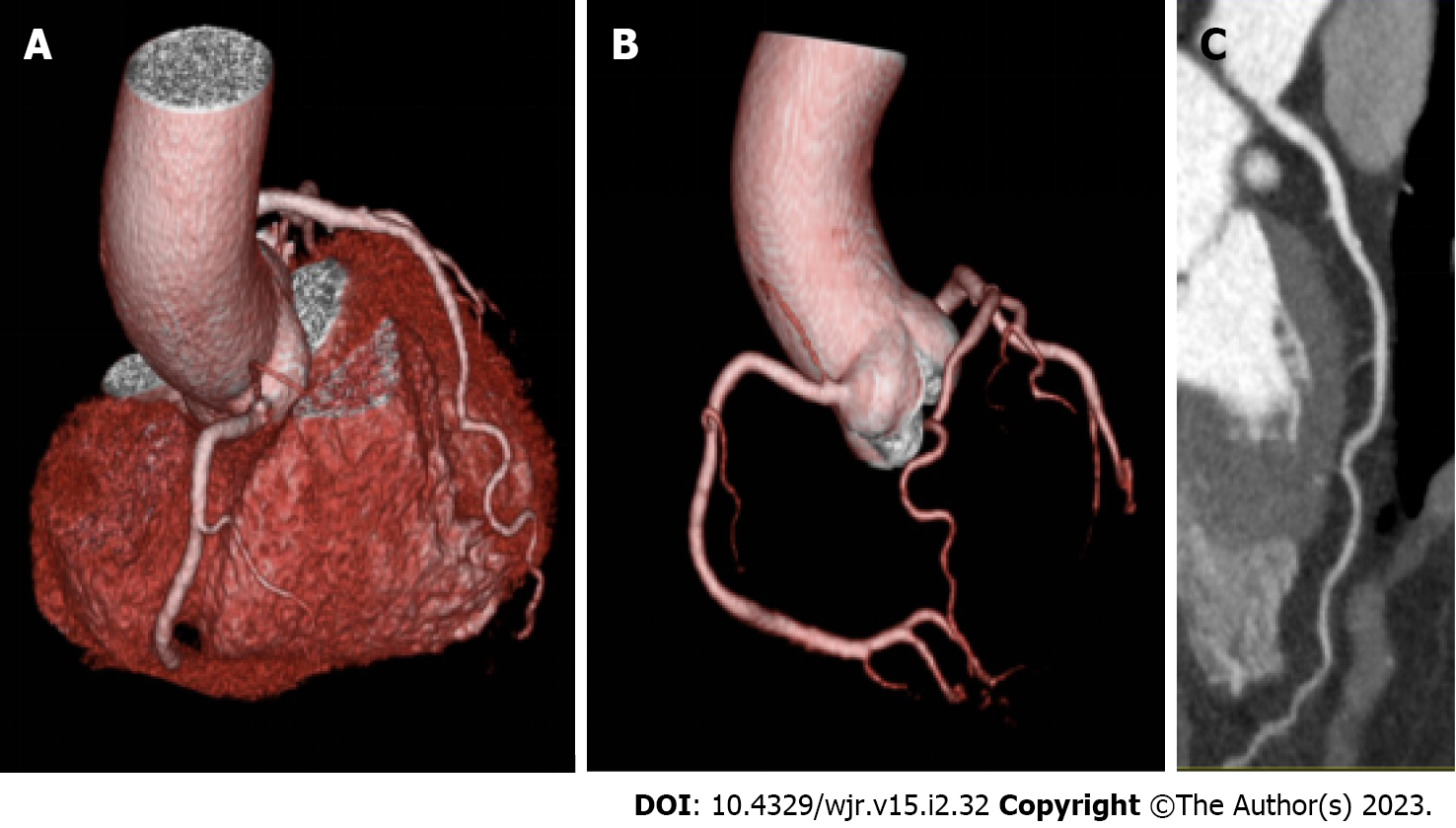
There was a total of 1603 potentially evaluable segments. Among these, in the 100 kV group, 40 segments were deemed unevaluable, and in the 80 kV group, 49 segments were deemed unevaluable due to having a diameter of < 1.5 mm or occlusion. The statistics for the coronary artery segments in groups A1 and A2 were presented in Table 2. The average IQ score for groups A1 and A2 was 4.46 ± 0.59 and 4.45 ± 0.62, respectively (Figures 3-5). There were no significant differences in IQ scores among the two groups (P equal to 0.08-0.31). According to the observers, the subjective IQ values of the two groups were very high (intraclass correlation coefficients equal to 0.71-0.90; Tables 3 and 4).
| Group | All segments | 5 | 4 | 3 | 2 | 1 | Average IQ score |
| A1 | 1085 | 420 (38.71%) | 611 (56.48%) | 45 (4.15%) | 6 (0.55%) | 1 (0.09%) | 4.46 ± 0.59 |
| A2 | 518 | 258 (49.81%) | 232 (44.79%) | 19 (3.67%) | 8 (1.54%) | 1 (0.19%) | 4.45 ± 0.62 |
| P value | 0.12 |
In the present study, the multiple dose reduction scan techniques significantly decreased the ED during the CCTA of patients under clinical diagnosis situations. The radiation dose for the group with multiple dose reduction was approximately 33.63% (1.13 ± 0.35/3.36 ± 1.30) of the dose associated with the conventional method, and the proportion of IQ that met the needs for the clinical diagnosis was > 99% (images with IQ scores of ≥ 3). There was no difference in subjective IQ when compared to conventional coronary CT scans.
Previous studies have only employed lower tube voltages, smart milliampere[1,7,14], or merely a narrow acquisition window for dose reduction techniques in CCTA[16,17]. To our knowledge, the present study was the first in the English literature to combine multiple technologies (narrow acquisition window, tube voltage of 80 kV, smart milliampere, and isocentric scanning) to reduce the CCTA radiation dose. The ED was significantly higher in the conventional group when compared to the multi-modality group (P < 0.05). A previous study performed a low-dose CCTA with the a CT scanner by Canon with 640 slices on patients whose heart rate was less than 70 beats/min and reported the mean ED as 2.67 ± 0.5 mSv[14]. Similar to this, using the same signal equipment (Aquilion ONE Genesis, Canon Medical Systems), Di Cesare et al[18] and Li et al[7] determined that ED medians were 2.80 ± 0.57 and 3.36 ± 2.35 mSv, respectively. During the present study, the value of the latter was the same as that of the conventional group. Although multi-modality technology was used to reduce the radiation dose of CCTA, there were no significant differences in subjective IQ scores (P = 0.12).
The present study revealed that the radiation dose of CCTA significantly decreased after the application of multiple dose reduction scan techniques. The radiation dose in group A1 was approximately 33.63% (1.13 ± 0.35/3.36 ± 1.30) in comparison with the traditional method. Low-dose CCTA has been shown to significantly reduce ED in some studies using dual source CTs (Siemens Healthcare)[7,14], GE gem energy spectrum CTs (GE Medical Systems)[8], as well as Philips Brilliance 256-slice iCTs (Philips Medical Systems)[4], with a tube voltage of 80 kV. The current research was the first to report the use of a low-dose CCTA at an 80 kV tube voltage utilizing a Canon CT system. A craniocaudal coverage of 16 cm was achieved, exceeding the length of the median scan employed in clinical practice. Furthermore, the tube generally rotates 360°, allowing it to be sufficient for cardiac scanning. Li et al[7] and Khosa et al[17] performed CCTA using the Canon 320-row dynamic CT system (Aquilion ONE, Toshiba Medical Systems; the present study used the same signal equipment), with a tube voltage of 120 kV. Both studies used the following parameters: RR interval, 66%-80%; BMI, 28 kg/m2; and heart rate, < 70 bpm. Furthermore, the present study employed 1.54 mSv, while the study conducted by Khosa et al[17] employed 633 mSv. According to some studies, radiation doses can reach 1.76 ± 0.43-2.72 ± 0.50 mSv when CCTA is used with an 80 kV tube voltage[7,13] when compared to the doses in the present study, and the ED was high.
A positive correlation exists between the intensity of X-rays and the square of tube voltage and current. In contrast to lowering tube current, reducing tube voltage substantially reduces radiation dose. In previous research, low tube voltage was found to reduce radiation dosage. However, this has the disadvantage of increasing picture noise and lowering CNR, which limits the reduction of the radiation dose[17-19]. Adaptive Iterative Dose Reduction Using Three Dimensional Processing was used in those studies to reconstruct the image, and the results were compared using conventional methods. This approach effectively improved the IQ and reduced the radiation dose in scans for different areas[17-19]. In another study, the use of Adaptive Iterative Dose Reduction Using Three Dimensional Processing and ultra-low-dose CT has been shown to significantly increase IQ[20].
Computed tomography angiography (CTA) is a non-invasive examination method and has the advantages of convenience, speed, safety, and reliability. This technique can clearly display the stenosis of the coronary lumen and plaque on the wall and determine whether the coronary artery has stenosis. Furthermore, its accuracy for CHD diagnosis is very high[17-19,21]. Digital subtraction angiography (DSA) is an invasive examination and is poorly accepted by patients. Coronary DSA can only determine vascular stenosis. Although coronary DSA is the gold standard for accurately determining vascular stenosis, it is an invasive examination with high radiation dose, high cost, and poor patient acceptance. A dose reduction on CTA examinations, such as the reduction of radiation exposure, could benefit patients with suspected CHD, who could not endure a DSA examination[22]. Our research group is performing further explorations on the effects of dose reduction techniques for patients who could not endure DSA examinations in order to increase its clinical practicability.
There are a few limitations to the current research. A minimum heart rate less than 70 beats/min was required for all patients, including those with low BMIs. A second drawback is that there was no correlation between the CCTA and DSA for the coronary artery, and the IQ was only rated subjectively for the coronary artery. A more in-depth investigation is required for individuals who are classified as obese according to their obesity grade (grade 1, BMI = 30-37.5 kg/m2 and grade 3, BMI ≥ 37.5 kg/m2).
Multiple dose reduction scan techniques can significantly reduce the radiation dose under conditions that meet the requirements for clinical diagnosis.
Coronary computed tomography angiography (CCTA) is considered to be an ideal non-invasive test for coronary heart disease (CHD). Nevertheless, as more people become aware of the dangers associated with radiation, the issue of computed tomography radiation has emerged as a major concern.
The present study aimed to explore the value of multiple dose reduction techniques on CCTA.
A consecutive sample of individuals with clinically suspected CHD was screened for inclusion in the current research. The inclusion criteria were: ≥ 18-years-old; heart rate less than 70 beats/min; and body mass index of 18.0 to 29.9 kg/m2. Patients having pacemakers, significant respiratory artifacts, metallic implants located within the scanning range, an allergy to contrast agents or Betaloc, or patients who had undergone cardiac surgery or had a history of cardiac tumors were not eligible for treatment.
Consecutive normal and overweight patients were prospectively divided into two groups: Group A1, patients who received multiple dose reduction scans (n = 82); and group A2, patients who received conventional scans (n = 39). The scan parameters for group A1 consisted of the following: An isocentric scan, a tube voltage of 80 kV, and an 80% smart milliampere for tube current control. The scan parameters for group A2 were as follows: Normal position, tube voltage = 100 kV, and smart mill
The average effective doses (EDs) for groups A1 and A2 were 1.13 ± 0.35 and 3.36 ± 1.30 mSv, respectively. A statistically significant difference was found between the two groups in terms of ED (P < 0.01). The signal-to-noise ratio as well as contrast signal-to-noise ratio of group A2 were significantly higher than those of group A1, in addition to having less noise. Moreover, the subjective image quality (IQ) scores were excellent in both groups, and the subjective IQ scores of the two groups did not differ significantly (P = 0.12).
Multiple dose reduction scan techniques can significantly decrease the ED of patients receiving CCTA examinations for clinical diagnosis.
This study supports the application of the multiple dose reduction scan techniques in patients receiving CCTA.
Provenance and peer review: Unsolicited article; Externally peer reviewed.
Peer-review model: Single blind
Specialty type: Radiology, nuclear medicine and medical imaging
Country/Territory of origin: China
Peer-review report’s scientific quality classification
Grade A (Excellent): 0
Grade B (Very good): B
Grade C (Good): C
Grade D (Fair): D
Grade E (Poor): 0
P-Reviewer: Crocé LS, Italy; El-Serafy AS, Egypt; Shariati MBH, Iran S-Editor: Fan JR L-Editor: Filipodia P-Editor: Fan JR
| 1. | Jia CF, Zhong J, Meng XY, Sun XX, Yang ZQ, Zou YJ, Wang XY, Pan S, Yin D, Wang ZQ. Image quality and diagnostic value of ultra low-voltage, ultra low-contrast coronary CT angiography. Eur Radiol. 2019;29:3678-3685. [RCA] [PubMed] [DOI] [Full Text] [Cited by in Crossref: 7] [Cited by in RCA: 5] [Article Influence: 0.8] [Reference Citation Analysis (0)] |
| 2. | Serruys PW, Hara H, Garg S, Kawashima H, Nørgaard BL, Dweck MR, Bax JJ, Knuuti J, Nieman K, Leipsic JA, Mushtaq S, Andreini D, Onuma Y. Coronary Computed Tomographic Angiography for Complete Assessment of Coronary Artery Disease: JACC State-of-the-Art Review. J Am Coll Cardiol. 2021;78:713-736. [RCA] [PubMed] [DOI] [Full Text] [Cited by in Crossref: 10] [Cited by in RCA: 97] [Article Influence: 24.3] [Reference Citation Analysis (0)] |
| 3. | Aghayev A, Murphy DJ, Keraliya AR, Steigner ML. Recent developments in the use of computed tomography scanners in coronary artery imaging. Expert Rev Med Devices. 2016;13:545-553. [RCA] [PubMed] [DOI] [Full Text] [Cited by in Crossref: 18] [Cited by in RCA: 20] [Article Influence: 2.2] [Reference Citation Analysis (0)] |
| 4. | Tang S, Zhang G, Chen Z, Liu X, He L. Application of prospective ECG-gated multiphase scanning for coronary CT in children with different heart rates. Jpn J Radiol. 2021;39:946-955. [RCA] [PubMed] [DOI] [Full Text] [Cited by in RCA: 4] [Reference Citation Analysis (0)] |
| 5. | Apfaltrer G, Albrecht MH, Schoepf UJ, Duguay TM, De Cecco CN, Nance JW, De Santis D, Apfaltrer P, Eid MH, Eason CD, Thompson ZM, Bauer MJ, Varga-Szemes A, Jacobs BE, Sorantin E, Tesche C. High-pitch low-voltage CT coronary artery calcium scoring with tin filtration: accuracy and radiation dose reduction. Eur Radiol. 2018;28:3097-3104. [RCA] [PubMed] [DOI] [Full Text] [Cited by in Crossref: 20] [Cited by in RCA: 34] [Article Influence: 4.9] [Reference Citation Analysis (0)] |
| 6. | Zhang W, Ba Z, Wang Z, Lv H, Zhao J, Zhang Y, Zhang F, Song L. Diagnostic performance of low-radiation-dose and low-contrast-dose (double low-dose) coronary CT angiography for coronary artery stenosis. Medicine (Baltimore). 2018;97:e11798. [RCA] [PubMed] [DOI] [Full Text] [Full Text (PDF)] [Cited by in Crossref: 4] [Cited by in RCA: 5] [Article Influence: 0.7] [Reference Citation Analysis (0)] |
| 7. | Li RF, Hou CL, Zhou H, Dai YS, Jin LQ, Xi Q, Zhang JH. Comparison on radiation effective dose and image quality of right coronary artery on prospective ECG-gated method between 320 row CT and 2nd generation (128-slice) dual source CT. J Appl Clin Med Phys. 2020;21:256-262. [RCA] [PubMed] [DOI] [Full Text] [Full Text (PDF)] [Cited by in Crossref: 4] [Cited by in RCA: 6] [Article Influence: 1.2] [Reference Citation Analysis (0)] |
| 8. | Liu K, Diao K, Hu S, Xu X, Zhang J, Peng W, Xia C, Zhang K, Li Y, Guo Y, He S, He Y, Li Z. Achieving Low Radiation Dose in “One-Stop” Myocardial Computed Tomography Perfusion Imaging in Coronary Artery Disease Using 16-cm Wide Detector CT. Acad Radiol. 2020;27:1531-1539. [RCA] [PubMed] [DOI] [Full Text] [Cited by in Crossref: 1] [Cited by in RCA: 1] [Article Influence: 0.2] [Reference Citation Analysis (0)] |
| 9. | Ali I, Ahmad S. Evaluation of the effects of sagging shifts on isocenter accuracy and image quality of cone-beam CT from kV on-board imagers. J Appl Clin Med Phys. 2009;10:180-194. [RCA] [PubMed] [DOI] [Full Text] [Full Text (PDF)] [Cited by in Crossref: 22] [Cited by in RCA: 17] [Article Influence: 1.1] [Reference Citation Analysis (0)] |
| 10. | Chow JC. Cone-beam CT dosimetry for the positional variation in isocenter: a Monte Carlo study. Med Phys. 2009;36:3512-3520. [RCA] [PubMed] [DOI] [Full Text] [Cited by in Crossref: 11] [Cited by in RCA: 12] [Article Influence: 0.8] [Reference Citation Analysis (0)] |
| 11. | Dane B, O’Donnell T, Liu S, Vega E, Mohammed S, Singh V, Kapoor A, Megibow A. Radiation dose reduction, improved isocenter accuracy and CT scan time savings with automatic patient positioning by a 3D camera. Eur J Radiol. 2021;136:109537. [RCA] [PubMed] [DOI] [Full Text] [Cited by in Crossref: 8] [Cited by in RCA: 9] [Article Influence: 2.3] [Reference Citation Analysis (0)] |
| 12. | Olden KL, Kavanagh RG, James K, Twomey M, Moloney F, Moore N, Carey K, Murphy KP, Grey TM, Nicholson P, Chopra R, Maher MM, O’Connor OJ. Assessment of isocenter alignment during CT colonography: Implications for clinical practice. Radiography (Lond). 2018;24:334-339. [RCA] [PubMed] [DOI] [Full Text] [Cited by in Crossref: 4] [Cited by in RCA: 4] [Article Influence: 0.6] [Reference Citation Analysis (0)] |
| 13. | Khan AR, Awan FR, Najam SS, Islam M, Siddique T, Zain M. Elevated serum level of human alkaline phosphatase in obesity. J Pak Med Assoc. 2015;65:1182-1185. [PubMed] |
| 14. | Cao JX, Wang YM, Lu JG, Zhang Y, Wang P, Yang C. Radiation and contrast agent doses reductions by using 80-kV tube voltage in coronary computed tomographic angiography: a comparative study. Eur J Radiol. 2014;83:309-314. [RCA] [PubMed] [DOI] [Full Text] [Cited by in Crossref: 35] [Cited by in RCA: 40] [Article Influence: 3.6] [Reference Citation Analysis (0)] |
| 15. | Nakanishi R, Sankaran S, Grady L, Malpeso J, Yousfi R, Osawa K, Ceponiene I, Nazarat N, Rahmani S, Kissel K, Jayawardena E, Dailing C, Zarins C, Koo BK, Min JK, Taylor CA, Budoff MJ. Automated estimation of image quality for coronary computed tomographic angiography using machine learning. Eur Radiol. 2018;28:4018-4026. [RCA] [PubMed] [DOI] [Full Text] [Cited by in Crossref: 13] [Cited by in RCA: 19] [Article Influence: 2.7] [Reference Citation Analysis (0)] |
| 16. | Smit EB, Bouwstra H, Roorda LD, van der Wouden JHC, Wattel ELM, Hertogh CMPM, Terwee CB. A Patient-Reported Outcomes Measurement Information System Short Form for Measuring Physical Function During Geriatric Rehabilitation: Test-Retest Reliability, Construct Validity, Responsiveness, and Interpretability. J Am Med Dir Assoc. 2021;22:1627-1632.e1. [RCA] [PubMed] [DOI] [Full Text] [Cited by in Crossref: 3] [Cited by in RCA: 10] [Article Influence: 2.5] [Reference Citation Analysis (0)] |
| 17. | Khosa F, Khan A, Nasir K, Shuaib W, Budoff M, Blankstein R, Clouse ME. Influence of image acquisition on radiation dose and image quality: full vs narrow phase window acquisition using 320 MDCT. ScientificWorldJournal. 2013;2013:731590. [RCA] [PubMed] [DOI] [Full Text] [Full Text (PDF)] [Cited by in Crossref: 5] [Cited by in RCA: 5] [Article Influence: 0.5] [Reference Citation Analysis (0)] |
| 18. | Di Cesare E, Gennarelli A, Di Sibio A, Felli V, Splendiani A, Gravina GL, Masciocchi C. Image quality and radiation dose of single heartbeat 640-slice coronary CT angiography: a comparison between patients with chronic atrial fibrillation and subjects in normal sinus rhythm by propensity analysis. Eur J Radiol. 2015;84:631-636. [RCA] [PubMed] [DOI] [Full Text] [Cited by in Crossref: 29] [Cited by in RCA: 34] [Article Influence: 3.4] [Reference Citation Analysis (0)] |
| 19. | Liu S, Fu Q, Yu H, Zhang C, Yang Q, Hu Y, Xu W. Evaluate of the effect of low tube voltage on the radiation dosage using 640-slice coronary CT angiography. J Xray Sci Technol. 2018;26:463-471. [RCA] [PubMed] [DOI] [Full Text] [Cited by in Crossref: 2] [Cited by in RCA: 2] [Article Influence: 0.3] [Reference Citation Analysis (0)] |
| 20. | Xiao M, Zhang M, Lei M, Hu X, Wang Q, Chen Y, Ye J, Xu R, Chen J. Application of ultra-low-dose CT in 3D printing of distal radial fractures. Eur J Radiol. 2021;135:109488. [RCA] [PubMed] [DOI] [Full Text] [Cited by in Crossref: 3] [Cited by in RCA: 8] [Article Influence: 1.6] [Reference Citation Analysis (1)] |
| 21. | Di Cesare E, Gennarelli A, Di Sibio A, Felli V, Perri M, Splendiani A, Gravina GL, Barile A, Masciocchi C. 320-row coronary computed tomography angiography (CCTA) with automatic exposure control (AEC): effect of 100 kV vs 120 kV on image quality and dose exposure. Radiol Med. 2016;121:618-625. [RCA] [PubMed] [DOI] [Full Text] [Cited by in Crossref: 10] [Cited by in RCA: 11] [Article Influence: 1.2] [Reference Citation Analysis (0)] |
| 22. | Guo W, Liu X, Gao Z, Pirbhulal S, Huang W, Lin WH, Zhang H, Tan N, Zhang YT. Quantification of three-dimensional computed tomography angiography for evaluating coronary luminal stenosis using digital subtraction angiography as the standard of reference. Biomed Eng Online. 2015;14:50. [RCA] [PubMed] [DOI] [Full Text] [Full Text (PDF)] [Cited by in Crossref: 6] [Cited by in RCA: 9] [Article Influence: 0.9] [Reference Citation Analysis (0)] |


