Copyright
©2014 Baishideng Publishing Group Inc.
World J Radiol. Aug 28, 2014; 6(8): 583-588
Published online Aug 28, 2014. doi: 10.4329/wjr.v6.i8.583
Published online Aug 28, 2014. doi: 10.4329/wjr.v6.i8.583
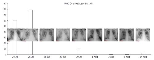
Figure 1 Correlation between white blood cell count and chest X-ray.
Note that the worsening of chest X-ray is directly linked with the fall of white blood cell due to the massive differentiation during the onset of differentiation syndrome. References: Institute of Radiology, Department of Clinical and Biological Sciences of University of Turin, AOU S. Luigi Gonzaga, Regione Gonzole 10, 10043 Orbassano, Torino, Italy. WBC: White blood cells.
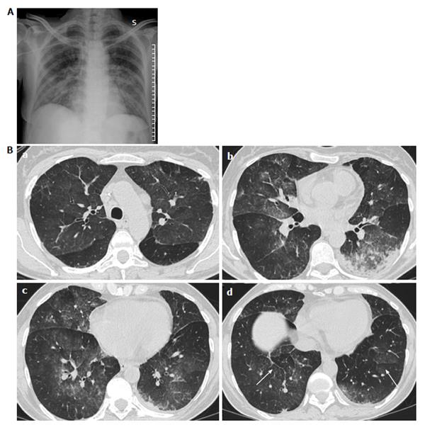
Figure 2 Photograph.
A: Chest X-ray shows subtile patchy ground glass opacities of the middle inferior lung fields; B: Computed tomography scans of the same patient as Figure 4 shows patchy ground glass opacities (a, b and c) with interlobar septal thickening (arrows in d). References: Institute of Radiology, Department of Clinical and Biological Sciences of University of Turin, AOU S.Luigi Gonzaga, Regione Gonzole 10, 10043 Orbassano, Torino, Italy.
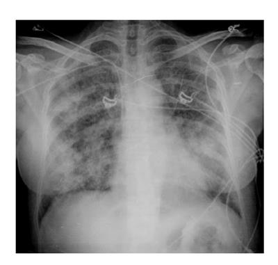
Figure 3 Supine Chest X-ray showing bilateral, asymmetrical patchy consolidation.
Septal lines and pleural effusions are absent. Based on the only radiologic features, it would be difficult to differentiate one fromacute respiratory distress syndrome or hemorrhage on these findings. References: Institute of Radiology, Department of Clinical and Biological Sciences of University of Turin, AOU S.Luigi Gonzaga, Regione Gonzole 10, 10043 Orbassano, Torino, Italy.
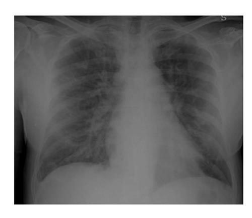
Figure 4 Chest X-ray in patient with differentiation syndrome showed mild cardiomegaly and increased pulmonary vascular marking in both lungs with thickening of small fissure and minimal pleural effusion on the right side seen as obliteration of right costo-phrenic angle: These findings are similar to those of congestive heart failure with pulmonary edema.
In contrast with congestive heart failure the time for the complete healing is long, and is similar to that of interstitial pneumonia or acute respiratory distress syndrome. References: Institute of Radiology, Department of Clinical and Biological Sciences of University of Turin, AOU S.Luigi Gonzaga, Regione Gonzole 10, 10043 Orbassano, Torino, Italy.
- Citation: Cardinale L, Asteggiano F, Moretti F, Torre F, Ulisciani S, Fava C, Rege-Cambrin G. Pathophysiology, clinical features and radiological findings of differentiation syndrome/all-trans-retinoic acid syndrome. World J Radiol 2014; 6(8): 583-588
- URL: https://www.wjgnet.com/1949-8470/full/v6/i8/583.htm
- DOI: https://dx.doi.org/10.4329/wjr.v6.i8.583


