Copyright
©2014 Baishideng Publishing Group Inc.
World J Radiol. Dec 28, 2014; 6(12): 890-894
Published online Dec 28, 2014. doi: 10.4329/wjr.v6.i12.890
Published online Dec 28, 2014. doi: 10.4329/wjr.v6.i12.890
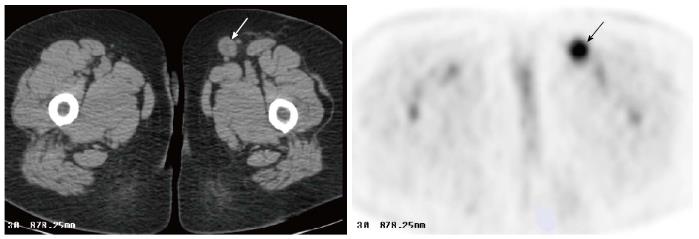
Figure 1 Axial image of F18-fluoro-2-deoxy-D-glucose positron emission tomography/computed tomography obtained 3 mo postoperatively in a 55-year-old woman with history of T2aN0Mx myxofibrosarcoma of the left ankle.
Compared to preoperative image, there was a new 2.3 cm left inguinal lymph node with intense uptake (SUV 8.0, arrows), suspicious for nodal metastasis. Biopsy of the node revealed reactive lymphadenitis. SUV: Standardized uptake value.
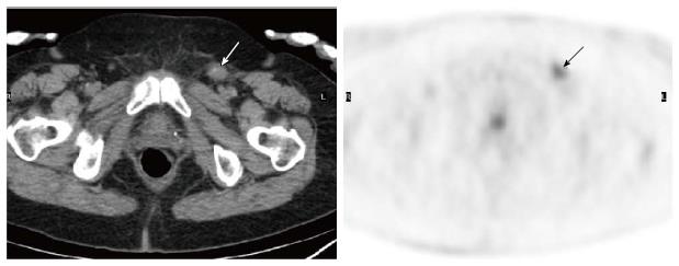
Figure 2 Axial image of F18-fluoro-2-deoxy-D-glucose positron emission tomography/computed tomography obtained 5 mo postoperatively in a 68-year-old woman with history of vulvar squamous cell carcinoma.
Compared to preoperative image, there was a new 1.5 cm left inguinal lymph node with increased uptake (SUV 4.9, arrows). Incisional biopsy of the node suggested lymphadenitis. SUV: Standardized uptake value.
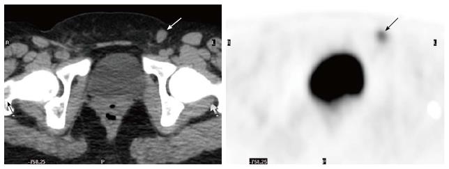
Figure 3 Axial image of F18-fluoro-2-deoxy-D-glucose positron emission tomography/computed tomography obtained 6 mo postoperatively in a 16-year-old woman with alveolar soft tissue sarcoma of the left knee.
Compared to preoperative image, there was a new 1.4 cm left inguinal lymph node with intense uptake (SUV 5.2, arrows). Incisional biopsy confirmed lymphadenitis. SUV: Standardized uptake value.
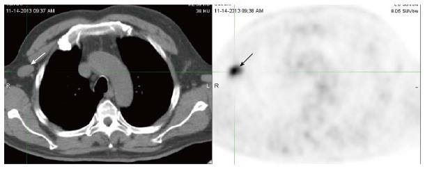
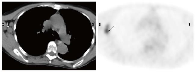
Figure 4 Axial image of F18-fluoro-2-deoxy-D-glucose positron emission tomography/computed tomography obtained 6 mo postoperatively in a 60-year-old man had history of tongue cancer.
The image demonstrated a 2.6 cm x 1.5 cm right axillary lymph node with intense uptake (SUV 6.1, arrows), suspicious for metastasis. Biopsy of the node suggested chronic lymphadenitis with reactive lymphoid hyperplasia. SUV: Standardized uptake value.
Figure 5 Axila image of F18-fluoro-2-deoxy-D-glucose positron emission tomography/computed tomography obtained 3 mo postoperatively in a 77-year-old woman had history of right breast cancer.
There were a few FDG avid right axillary lymph nodes, the largest 1.6 cm with SUV 5.1 (arrows), all new compared to prior images. The findings were highly suspicious for regional nodal metastases. Subsequent biopsy was indicative of reactive lymphadenitis. SUV: Standardized uptake value.
- Citation: Liu Y. Postoperative reactive lymphadenitis: A potential cause of false-positive FDG PET/CT. World J Radiol 2014; 6(12): 890-894
- URL: https://www.wjgnet.com/1949-8470/full/v6/i12/890.htm
- DOI: https://dx.doi.org/10.4329/wjr.v6.i12.890


