Copyright
©The Author(s) 2024.
World J Radiol. Sep 28, 2024; 16(9): 460-465
Published online Sep 28, 2024. doi: 10.4329/wjr.v16.i9.460
Published online Sep 28, 2024. doi: 10.4329/wjr.v16.i9.460
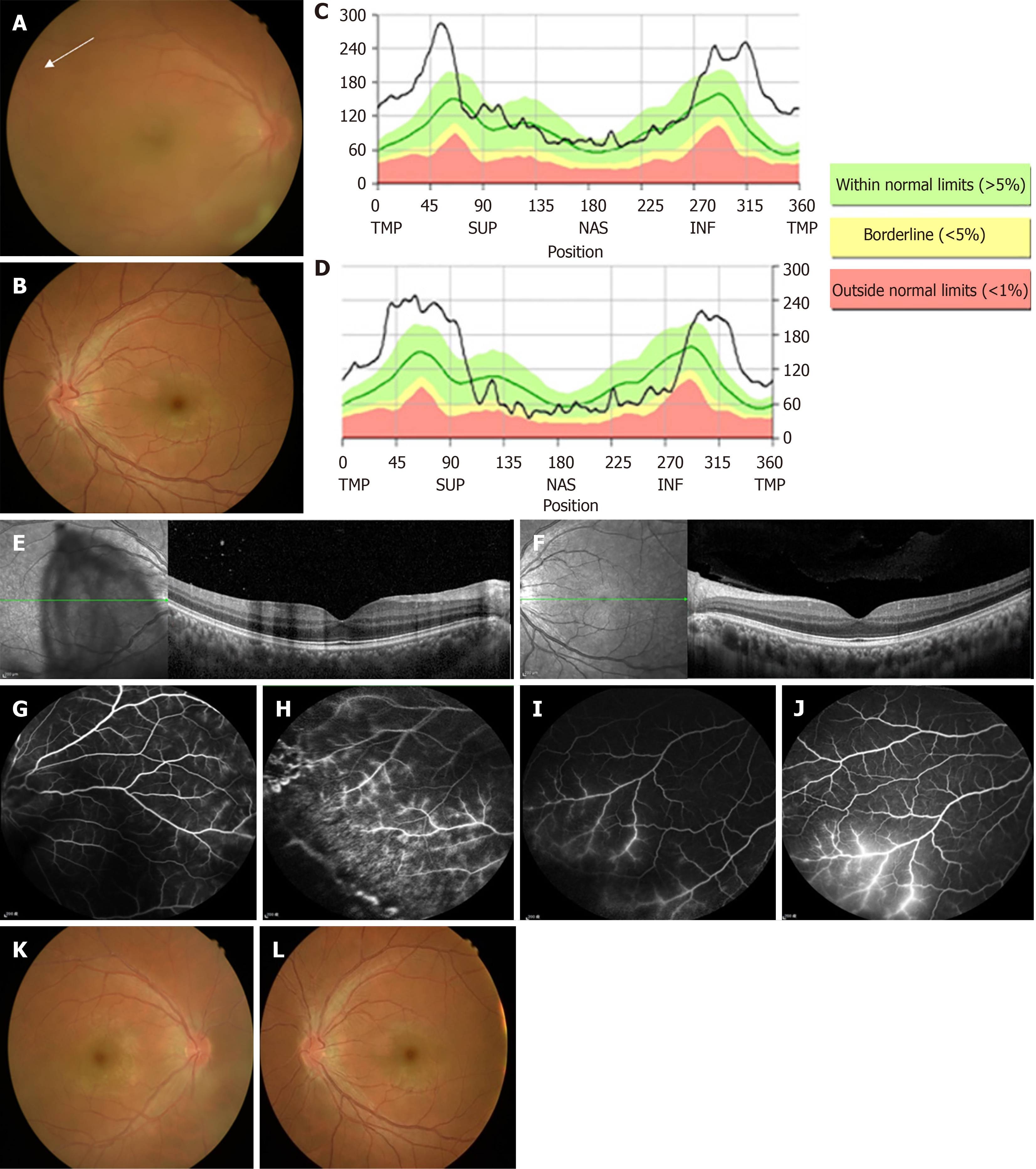
Figure 1 Fundus examination.
A and B: Fundus photographs revealing disc congestion, venous engorgement, and retinal hemorrhage at periphery (white arrow) in right eye (A) and left eye (B); C-F: Optical coherence tomography showed prominent bilateral disc edema (C and D) and increased inner retina thickness of macula in his right eye (E). The thickness of the left macula was unremarkable (F); G-J: Fluorescein angiography disclosed predominant phlebitis with perivascular staining at periphery in right eye (G and H) and left eye (I and J); K and L: Two weeks after treatment, the fundus of both eyes had cleared up.
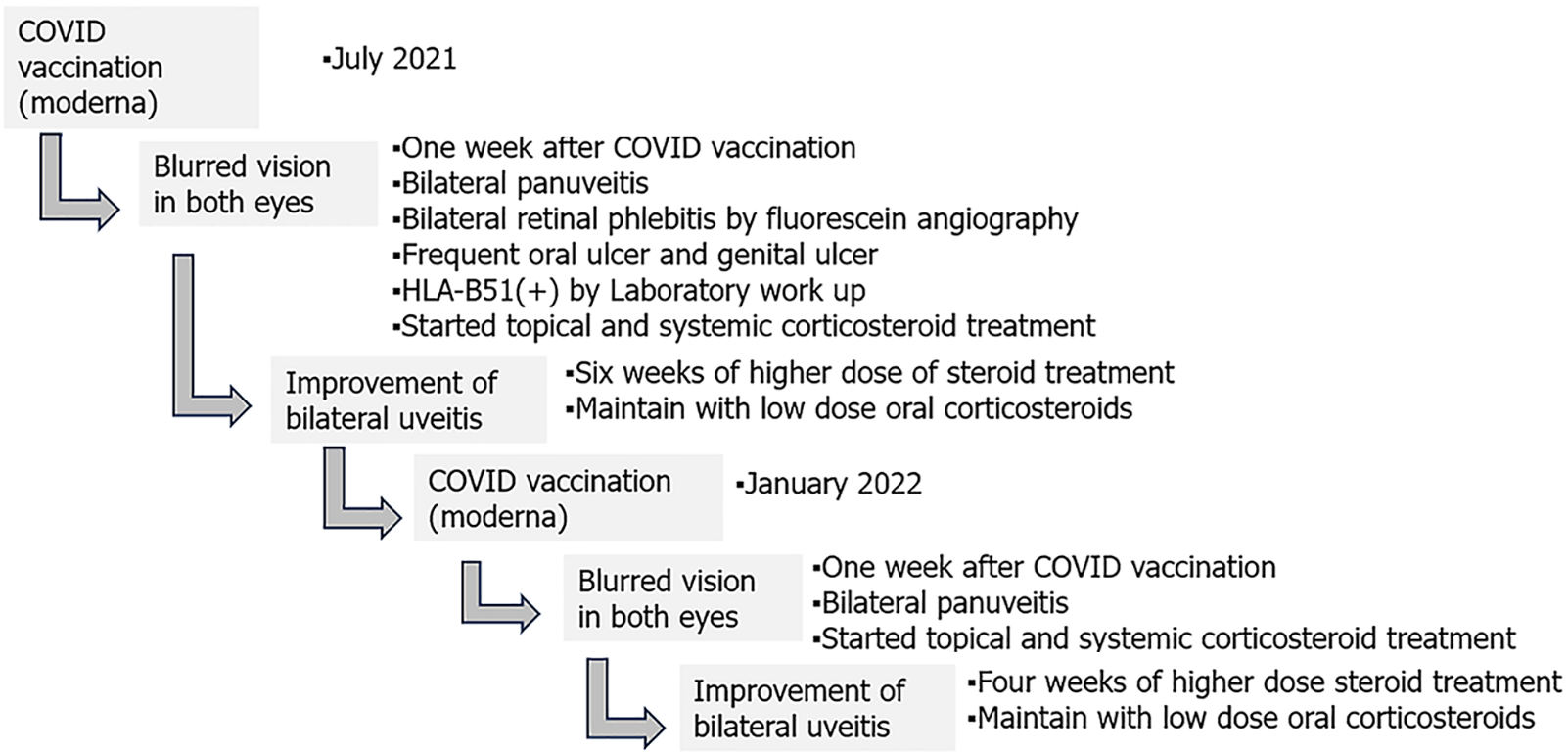
Figure 2 The patient’s timeline.
COVID: Coronavirus disease.
- Citation: Lin RT, Liu PK, Chang CW, Cheng KC, Chen KJ, Chang YC. Behcet's disease-related panuveitis following COVID-19 vaccination: A case report. World J Radiol 2024; 16(9): 460-465
- URL: https://www.wjgnet.com/1949-8470/full/v16/i9/460.htm
- DOI: https://dx.doi.org/10.4329/wjr.v16.i9.460


