Copyright
©2014 Baishideng Publishing Group Co.
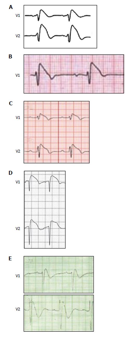
Figure 1 Type 1 Brugada phenocopies.
A: True congenital type 1 Brugada electrocardiogram (ECG) pattern; B: Type 1 Brugada phenocopies (BrP) in a patient with an acute inferior ST-segment elevation myocardial infarction with right ventricular involvement; C: Type 1 BrP in a patient with concurrent hyperkalemia, hyponatremia and acidosis; D: Type 1 BrP in a patient with an acute pulmonary embolism; E: Type 1 BrP in a patient with hypokalemia in the context of congenital hypokalemic periodic paralysis.
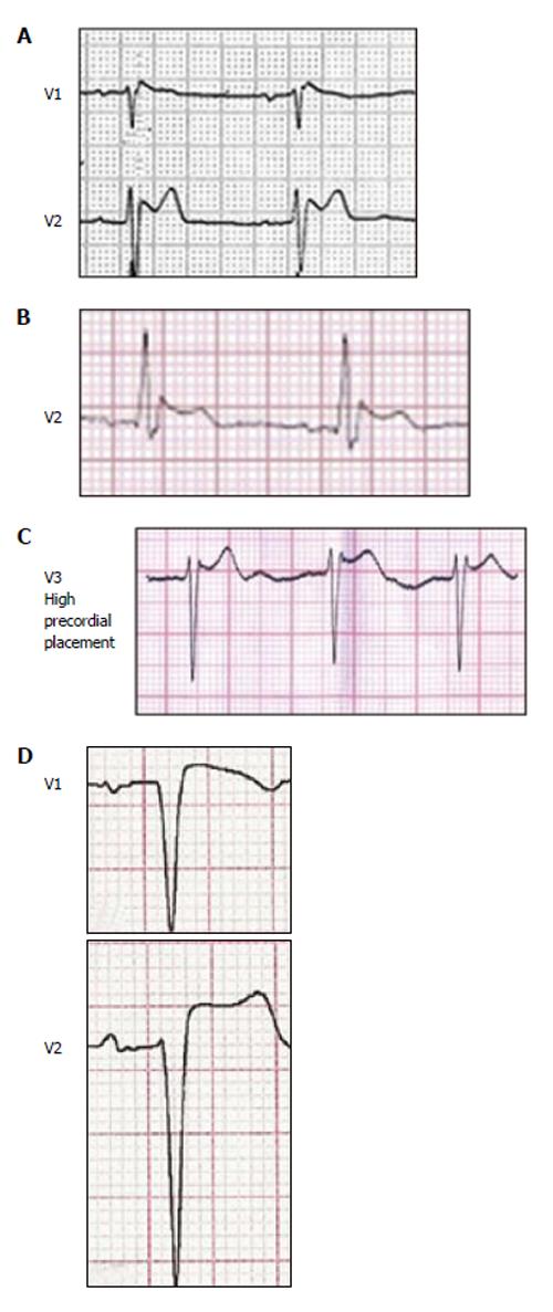
Figure 2 Type 2 Brugada phenocopies.
A: True congenital type 2 Brugada electrocardiogram (ECG) pattern; B: Type 2 Brugada phenocopies (BrP) in a patient with an accidental electrocution injury; C: Type 2 BrP in a patient with congenital pectus excavatum causing mechanical mediastinal compression; D: Type 2 BrP as a result of an inappropriate high pass ECG filter.
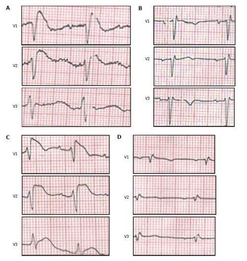
Figure 3 Brugada phenocopy clinical reproducibility.
A: Electrocardiogram (ECG) on presentation while the patient is hypokalemic consistent with a type 1 Brugada ECG pattern; B: After correction of the electrolyte abnormality, the ECG normalizes; C: While in hospital, the patient again becomes hypokalemic with recurrence of the type 1 Brugada ECG pattern; D: Subsequent normalization of the ECG pattern after potassium is corrected.
- Citation: Anselm DD, Evans JM, Baranchuk A. Brugada phenocopy: A new electrocardiogram phenomenon. World J Cardiol 2014; 6(3): 81-86
- URL: https://www.wjgnet.com/1949-8462/full/v6/i3/81.htm
- DOI: https://dx.doi.org/10.4330/wjc.v6.i3.81


