Copyright
©The Author(s) 2020.
World J Cardiol. Dec 26, 2020; 12(12): 599-614
Published online Dec 26, 2020. doi: 10.4330/wjc.v12.i12.599
Published online Dec 26, 2020. doi: 10.4330/wjc.v12.i12.599
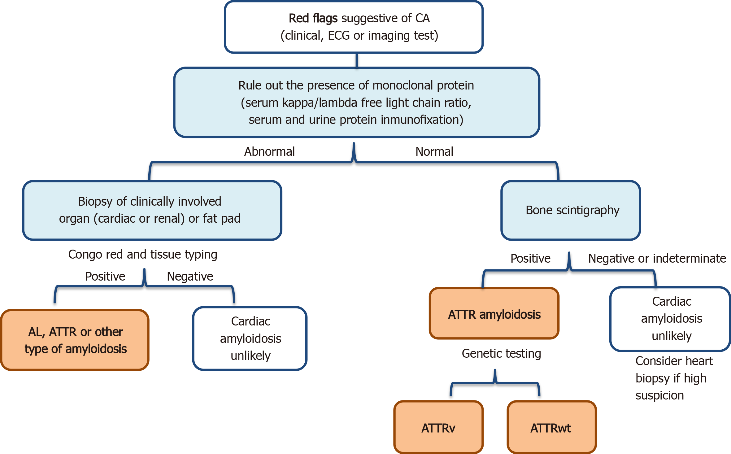
Figure 1 Algorithm for diagnostic patients with suspected cardiac amyloidosis[78].
AL: Light chain amyloidosis; ATTR: Transthyretin amyloidosis; ATTRv: Variant transthyretin amyloidosis; ATTRwt: Wild-type transthyretin amyloidosis; CA: Cardiac amyloidosis; ECG: Electrocardiogram.
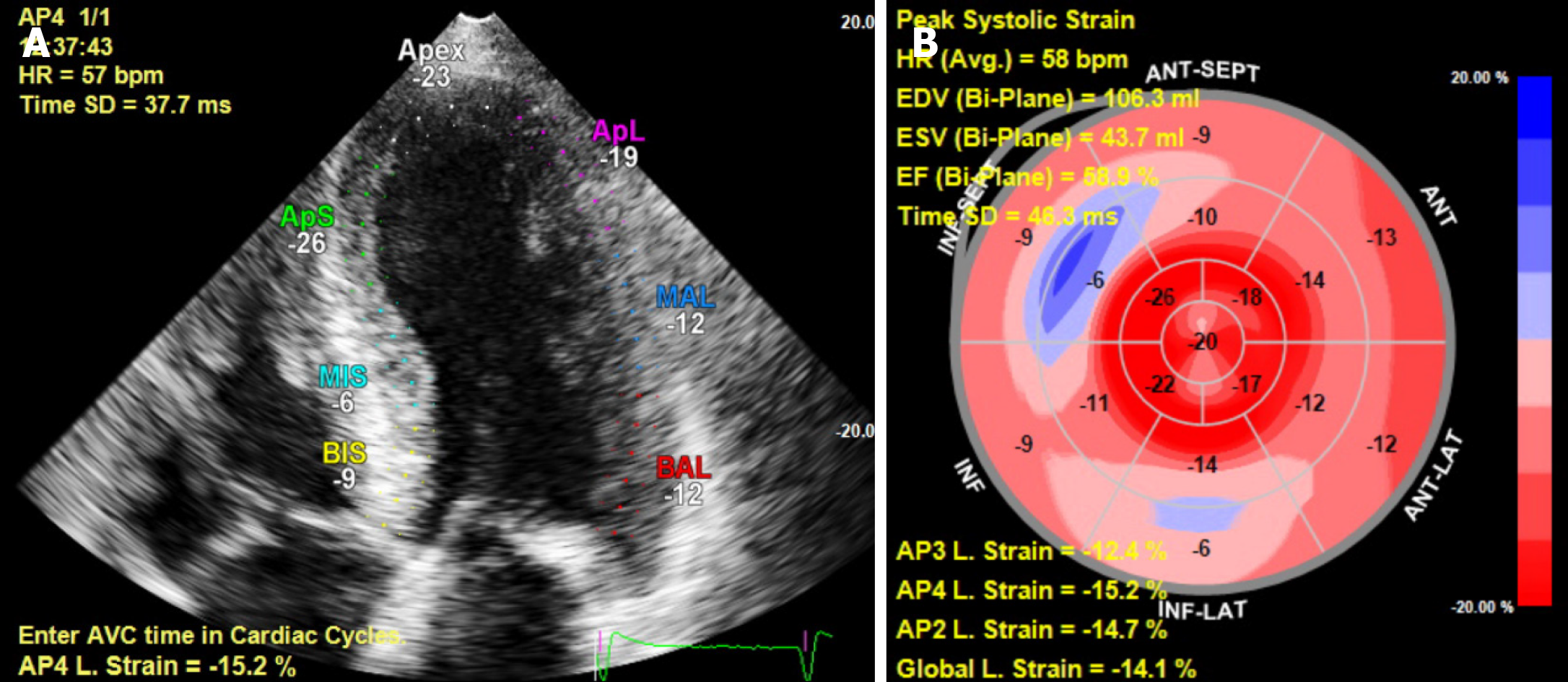
Figure 2 Transthoracic echocardiography imaging from a patient with transthyretin amyloidosis-cardiac amyloidosis.
A: Strain imaging by transthoracic echocardiography; B: Reduced mid and basal values with apical sparing.
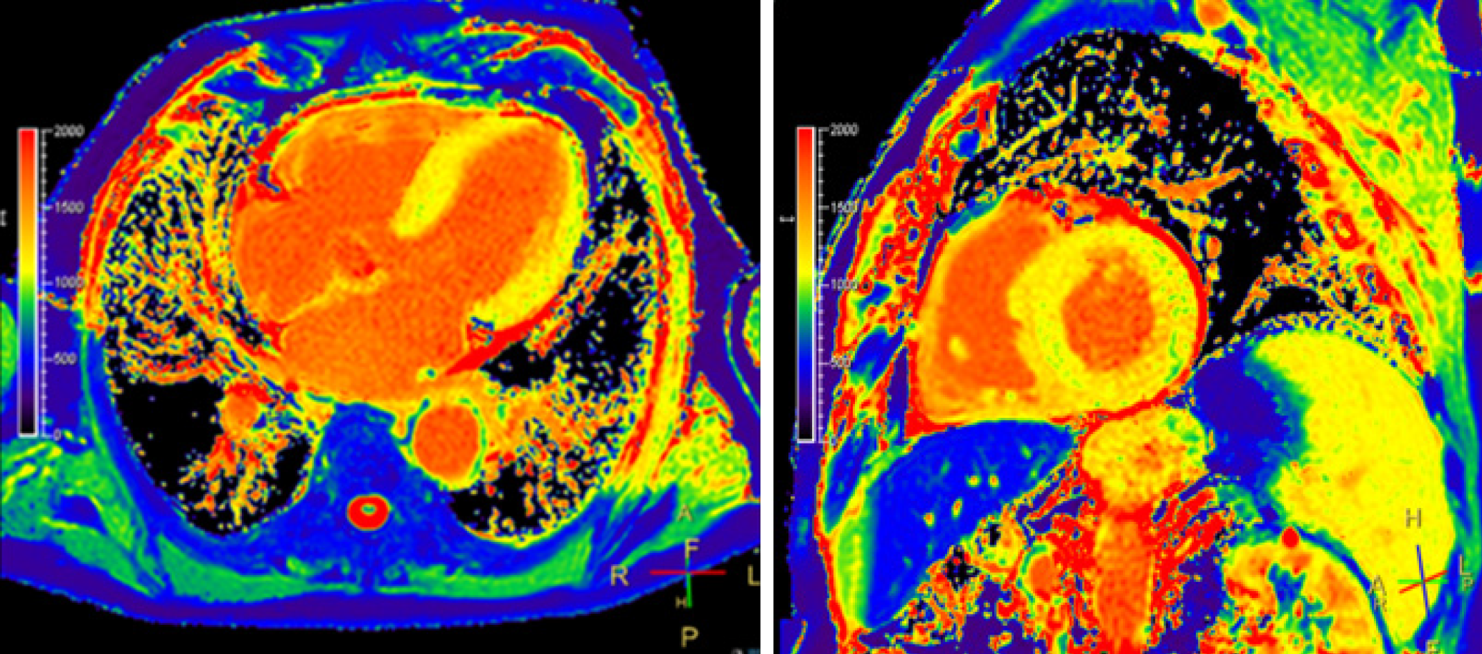
Figure 3 Native T1-mapping images.
Abnormally elevated values of T1 native (1120 ms) in a patient with amyloidosis-cardiac amyloidosis.
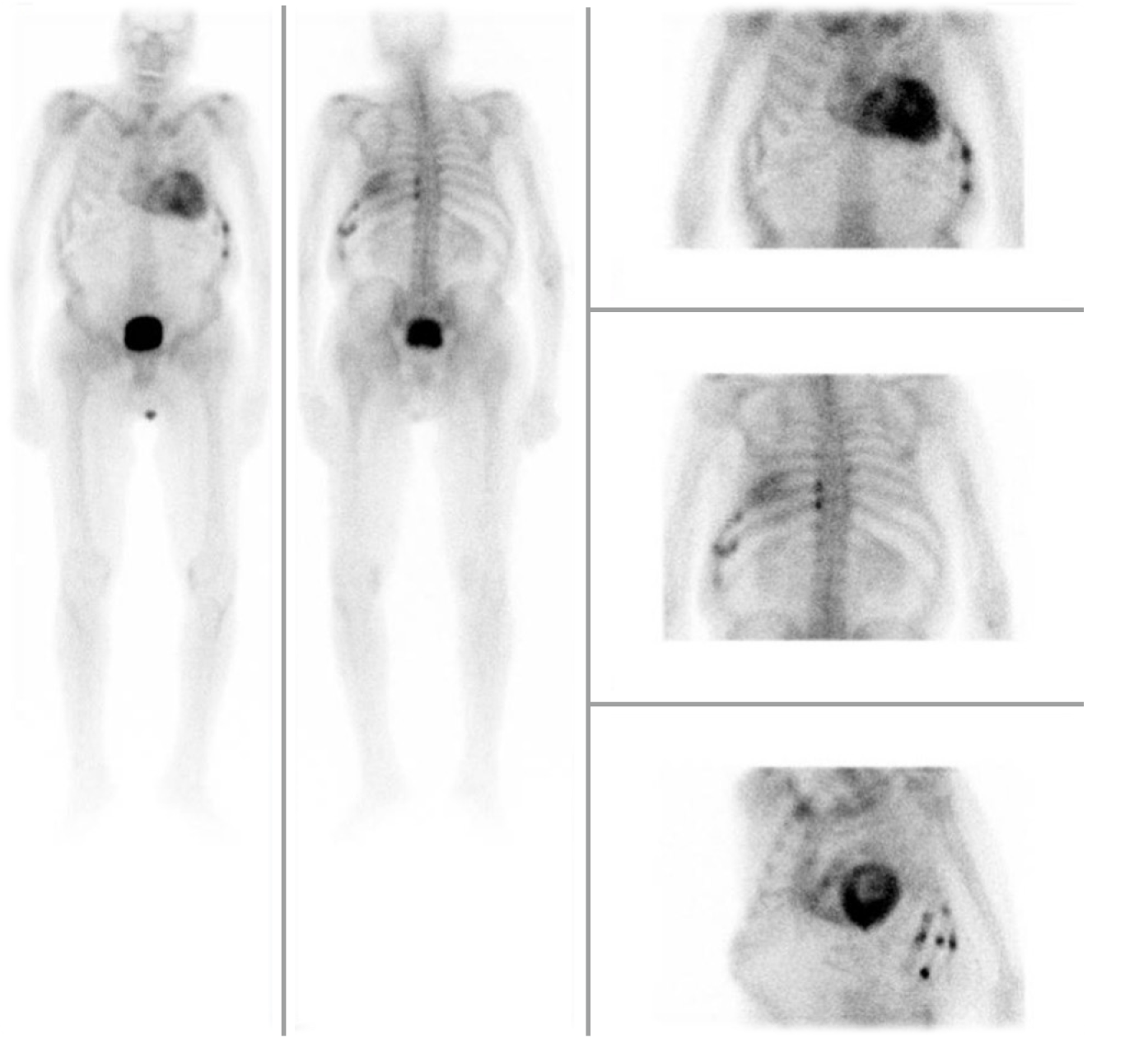
Figure 4 Planar scintigraphy showing intense cardiac uptake of 99mTc-labeled 3,3-diphosphono-1,2-propanodicarboxylic acid corresponding to a grade 3 in a patient diagnosed with cardiac amyloidosis-transthyretin amyloidosis.
- Citation: Vidal-Perez R, Vázquez-García R, Barge-Caballero G, Bouzas-Mosquera A, Soler-Fernandez R, Larrañaga-Moreira JM, Crespo-Leiro MG, Vazquez-Rodriguez JM. Diagnostic and prognostic value of cardiac imaging in amyloidosis. World J Cardiol 2020; 12(12): 599-614
- URL: https://www.wjgnet.com/1949-8462/full/v12/i12/599.htm
- DOI: https://dx.doi.org/10.4330/wjc.v12.i12.599


