Copyright
©The Author(s) 2017.
World J Biol Chem. Feb 26, 2017; 8(1): 32-44
Published online Feb 26, 2017. doi: 10.4331/wjbc.v8.i1.32
Published online Feb 26, 2017. doi: 10.4331/wjbc.v8.i1.32
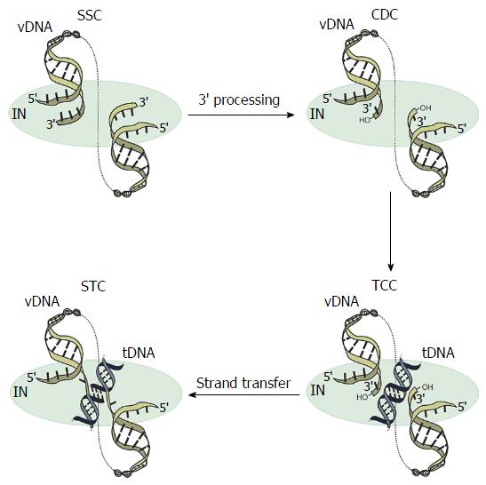
Figure 1 Integrase catalytic functions and intasome complexes.
A multimer of integrase (IN) (depicted simply by blue oval) engages the end regions of the linear vDNA molecule (yellow), forming the stable synaptic complex (SSC). During 3’-processing, IN hydrolyzes the vDNA ends adjacent to invariant CA dinucleotides, revealing a set of reactive 3’-hydroxyl groups in the confines of the cleaved donor complex (CDC). After nuclear localization, the target capture complex (TCC) is formed upon tDNA (black) capture. Strand transfer, whereby IN employs the 3’ hydroxyl groups as nucleophiles to attack the tDNA, marks the transition to the strand transfer complex (STC).
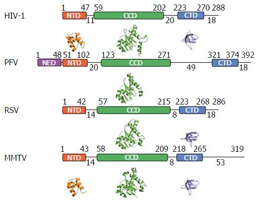
Figure 2 Integrase domain organization and representative secondary structures.
Starting and ending residues for integrase (IN) domains are indicated above the boxes, and interdomain linker lengths as well as C-terminal tail lengths are indicated below the lines. Crystal structures of N-terminal domains (NTDs), catalytic core domains (CCDs), and C-terminal domains (CTDs) are provided underneath the corresponding schematic IN representation. Crystal structures in the absence of DNA are not available for the PFV NTD extension domain (NED), NTD, or CTD, as well as for the RSV NTD. PDB accession codes: HIV-1 (NTD, 1K6Y; CCD, 1BIU; CTD, 1EX4), PFV (CCD, 3DLR), RSV (CCD, 1C0M; CTD, 1C0M) and MMTV (NTD, 5CZ2; CCD, 5CZ1; CTD, 5D7U). HIV: Human immunodeficiency virus; PFV: Prototype foamy virus; RSV: Rous sarcoma virus; MMTV: Mouse mammary tumor virus.
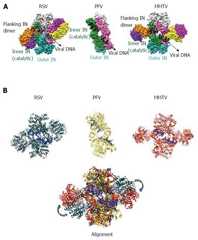
Figure 3 Comparison of prototype foamy virus, Rous sarcoma virus and mouse mammary tumor virus intasomes.
A: The PFV intasome comprises two catalytic inner subunits (green and pink) and two outer supportive INs (cyan and light grey). Only the CCDs of the outer subunits are discernable in crystallographic electron density maps. RSV and MMTV share the PFV intasome core architecture and employ two additional flanking IN dimers (orange-purple and yellow-dark pink) to complete the intasome structures; B: Three-dimensional alignment of RSV (grey, PDB accession code 5EJK), PFV (yellow, PDB code 3L2Q), and MMTV (red, PDB code 3JCA) intasome structures was performed using Chimera. For the MMTV intasome, flanking dimers were unambiguously positioned into the intasome core of the cryo-EM map via rigid-body docking. The alignment reveals a high degree of flexibility (approximately 30-40 Å) for the flanking RSV and MMTV dimers relative to the common intasome core structures (arrows). PFV: Prototype foamy virus; RSV: Rous sarcoma virus; MMTV: Mouse mammary tumor virus.
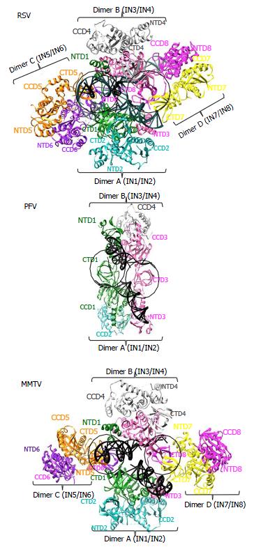
Figure 4 Integrase domain organizations within the prototype foamy virus, Rous sarcoma virus and mouse mammary tumor virus intasome structures.
Separate integrase (IN) domains are labeled, with IN monomer coloring code retained from Figure 3. The green IN1 and pink IN3 monomers donate their active sites for catalysis of 3’ processing and strand transfer across the structures. Circled areas represent similarly positioned CTDs. While these emanate from inner IN1 and IN3 monomers in the PFV structure, they originate from flanking MMTV and RSV IN monomers IN6 and IN8. PDB accession codes same as in Figure 3. PFV: Prototype foamy virus; RSV: Rous sarcoma virus; MMTV: Mouse mammary tumor virus; CCD: Catalytic core domain; NTD: N-terminal domain; CTD: C-terminal domain.
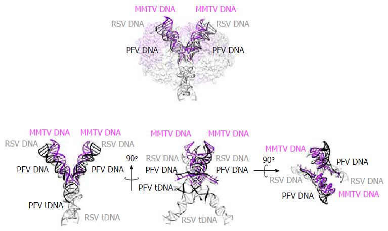
Figure 5 Superposition of prototype foamy virus strand transfer complex, Rous sarcoma virus strand transfer complex, and mouse mammary tumor virus cleaved donor complex structures with respect to vDNA and tDNA.
For simplicity, integrase content is either partially transparent or omitted. Color-coding is as following: PFV DNA: Black; RSV DNA: Grey; MMTV DNA: Purple. 90° rotations show different angles of the intasomes. PDB accession codes: PFV STC: 3OS0, RSV STC: 5EJK, MMTV CDC: 3JCA. PFV: Prototype foamy virus; RSV: Rous sarcoma virus; MMTV: Mouse mammary tumor virus; STC: Strand transfer complex; CDC: Cleaved donor complex.
- Citation: Grawenhoff J, Engelman AN. Retroviral integrase protein and intasome nucleoprotein complex structures. World J Biol Chem 2017; 8(1): 32-44
- URL: https://www.wjgnet.com/1949-8454/full/v8/i1/32.htm
- DOI: https://dx.doi.org/10.4331/wjbc.v8.i1.32


