Copyright
©2014 Baishideng Publishing Group Inc.
World J Biol Chem. Nov 26, 2014; 5(4): 398-408
Published online Nov 26, 2014. doi: 10.4331/wjbc.v5.i4.398
Published online Nov 26, 2014. doi: 10.4331/wjbc.v5.i4.398
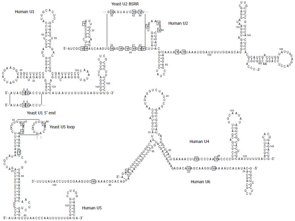
Figure 1 Primary sequences and secondary structures of human spliceosomal snRNAs (U1, U2, U4, U5 and U6).
Pseudouridines (Ψ) are boxed. The sequences of yeast snRNAs where the Ψs have their counterparts in human snRNAs (the 5’ end region of U1, branch site recognition region (BSRR) of U2, and loop region of U5) are also shown. The structures are predicted by the “multifold” program and are consistent with the genetic/biochemical mapping data.
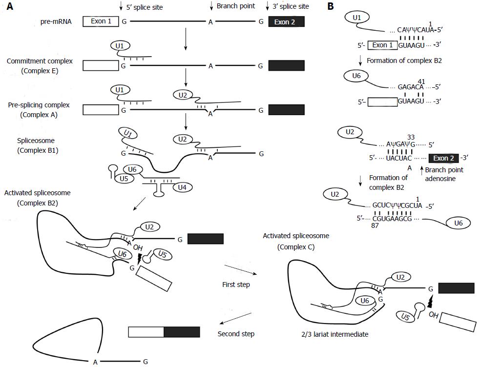
Figure 2 The splicing reaction.
A: Steps of the spliceosome-mediated splicing reaction. The thick lines represent the intron and the boxes are exons. The short lines between RNA strands represent watson-crick base-pairing interactions. The 2'-OH groups of branch point adenosine and the cut-off 5' exon are pictured in the activated spliceosomes. The lightning symbols depict a nucleophilic attack that causes a transesterification reaction; B: Putative RNA-RNA hybrids formed during the splicing reaction.
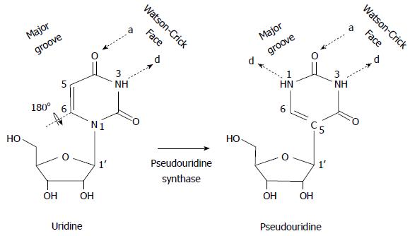
Figure 3 Schematic representation of uridine-to- pseudouridine isomerization.
Pseudouridine is a rotational isomer of uridine, in which the N-C glycosidic bond is broken to form the C-C bond. This results in the creation of an extra hydrogen bond donor (d), while the number of hydrogen bond acceptors (a) is unchanged.
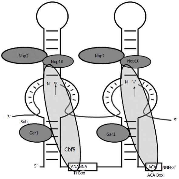
Figure 4 Schematic depiction of box H/ACA RNA.
The core components of a box H/ACA RNP, a box H/ACA RNA and four proteins (Nhp2, Nop 10, Gar1 and Cbf5), are shown. An RNA substrate paired with the two internal loops of the box H/ACA RNA is also shown. The arrows indicate the target nucleotides for pseudouridylation. The H box (5'-ANANNA-3') and ACA box (5'-ACA-3') are indicated.
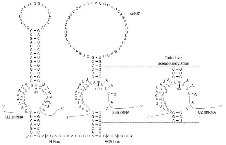
Figure 5 Constitutive and induced pseudouridylation by snR81 box H/ACA ribonucleoprotein.
The sequence and structure of snR81 box H/ACA RNA is shown. As arrows indicate, the internal loop (pseudouridylation pocket) within the 5’ hairpin is specific for Ψ42 (constitutive) of U2 snRNA, and the internal loop within the 3’ hairpin is specific for Ψ1051 (constitutive) of 25S rRNA. Under stress coditions, the 3’ pseudouridylation pocket becomes capable of directing the formation of Ψ93 (inducible) of U2 snRNA. As shown by “x”, there are two U-U mismatches between the 3’ pocket and the U2 sequence flanking position 93.
- Citation: Adachi H, Yu YT. Insight into the mechanisms and functions of spliceosomal snRNA pseudouridylation. World J Biol Chem 2014; 5(4): 398-408
- URL: https://www.wjgnet.com/1949-8454/full/v5/i4/398.htm
- DOI: https://dx.doi.org/10.4331/wjbc.v5.i4.398


