Copyright
©2012 Baishideng Publishing Group Co.
World J Gastrointest Surg. Jan 27, 2012; 4(1): 1-8
Published online Jan 27, 2012. doi: 10.4240/wjgs.v4.i1.1
Published online Jan 27, 2012. doi: 10.4240/wjgs.v4.i1.1
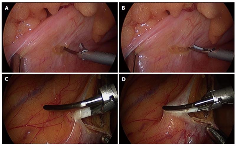
Figure 1 Retroperitoneum dissection.
A and B: After a small incision for the retroperitoneum is performed, the incision is enlarged, sealing forwards to the right while hooking with the ultrasonic surgical blade of the Harmonic ACE or clamping with the tissue pad and the ultrasonic surgical blade; C and D: When the incision is enlarged forwards to the left, cutting is performed by clamping with the ultrasonic surgical blade and the tissue pad of the Harmonic ACE, applying the tissue pad to the vessel side in order to decrease the thermal damage to adjacent structures.
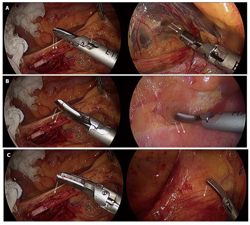
Figure 2 Dissection technique.
The left figures show the dissection face of the Harmonic ACE, whereas the right figures show that the Harmonic ACE is dissecting tissue. These methods are used by rounding the tip of the Harmonic ACE to various directions as needed, according to circumstances. A: A dissection using the sharp curve of the Harmonic ACE is shown. This method is used to dissect the tissue widely and quickly; B: A dissection by the round curve of the Harmonic ACE is shown. This is used to dissect the tissue carefully and gently; C: The blade tip of the Harmonic ACE is used for pinpoint dissection. These methods use rounding of the tip of the Harmonic ACE in various directions, according to the circumstances.
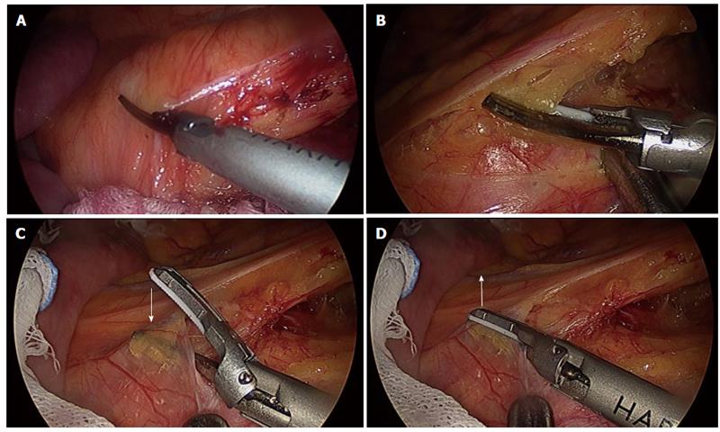
Figure 3 Dissection technique around the feeding artery.
A and B: The dissection around the feeding artery is enlarged by sealing while clamping with the ultrasonic surgical blade and the tissue pad of the Harmonic ACE, applying the tissue pad to the vessel side in order to decrease the thermal damage to adjacent structures; C and D: It is necessary to force the blade in the direction against the feeding artery when the ultrasonic surgical blade of the Harmonic ACE has to be applied to the feeding artery side; C shows the clamping with the ultrasonic surgical blade and the tissue pad of the Harmonic ACE; D shows the direction that must be forced with sealing.
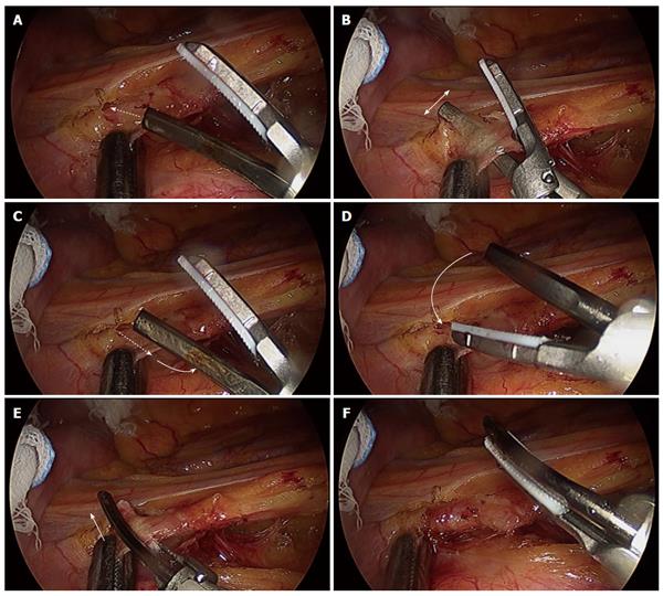
Figure 4 Skeletonization technique for the feeding artery.
A: First, the ultrasonic surgical blade of the Harmonic ACE penetrates the sheath of the feeding artery; B: The dissection margin is enlarged by moving the surgical blade along the feeding artery; C: After sufficient enlargement of the dissection margin for the sheath of the feeding artery, the ultrasonic surgical blade of the Harmonic ACE is removed from the sheath of the feeding artery; D: Next, the tip of the Harmonic ACE is rotated; E: The tissue pad side of the Harmonic ACE is applied in the penetrated space of the sheath of the feeding artery; F: One side of the sheath of the feeding artery is sealed and skeletonized safely. Then, the other side of the sheath is treated similarly, resulting in the complete skeletonization of the feeding artery.
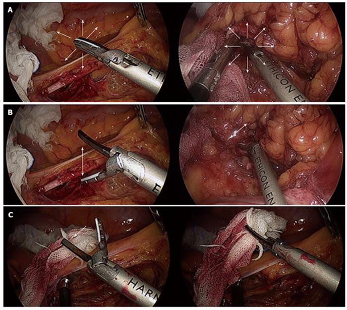
Figure 5 Various dissection techniques.
The left figures show the movement of the tip of the Harmonic ACE, whereas the right figures show that the Harmonic ACE is treating tissue. A: The dissection technique uses the closed tip of the Harmonic ACE as it is pushed, moving in the various directions; B: The Harmonic ACE can be used as a dissector, such as a Kelly forceps; C: The Harmonic ACE can be used as a grasper.
- Citation: Hotta T, Takifuji K, Yokoyama S, Matsuda K, Higashiguchi T, Tominaga T, Oku Y, Watanabe T, Nasu T, Hashimoto T, Tamura K, Ieda J, Yamamoto N, Iwamoto H, Yamaue H. Literature review of the energy sources for performing laparoscopic colorectal surgery. World J Gastrointest Surg 2012; 4(1): 1-8
- URL: https://www.wjgnet.com/1948-9366/full/v4/i1/1.htm
- DOI: https://dx.doi.org/10.4240/wjgs.v4.i1.1


