Copyright
©2011 Baishideng Publishing Group Co.
World J Gastrointest Surg. Oct 27, 2011; 3(10): 156-158
Published online Oct 27, 2011. doi: 10.4240/wjgs.v3.i10.156
Published online Oct 27, 2011. doi: 10.4240/wjgs.v3.i10.156
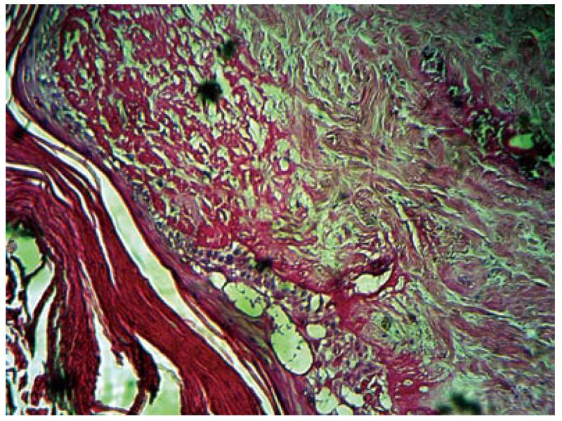
Figure 1 Histological examination with HE staining showed hyperkeratosis, atrophic epidermis, basal layer hydropic degeneration in epidermis, on dermis collagen deposition and subendothelial sclerosis in arterial wall in segmental foci that caused ischemic infarct leading to atrophy of adnexal structures.
- Citation: Ahmadi M, Rafi SA, Faham Z, Azhough R, Rooy SB, Rahmani O. A fatal case of Degos’ disease which presented with recurrent intestinal perforation. World J Gastrointest Surg 2011; 3(10): 156-158
- URL: https://www.wjgnet.com/1948-9366/full/v3/i10/156.htm
- DOI: https://dx.doi.org/10.4240/wjgs.v3.i10.156


