Copyright
©2010 Baishideng.
World J Gastrointest Surg. Apr 27, 2010; 2(4): 143-146
Published online Apr 27, 2010. doi: 10.4240/wjgs.v2.i4.143
Published online Apr 27, 2010. doi: 10.4240/wjgs.v2.i4.143
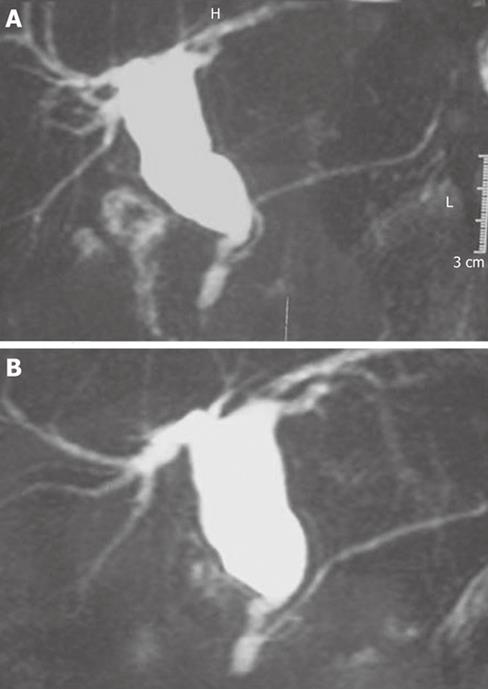
Figure 1 Magnetic resonance imaging.
A: Pancreaticobiliary maljunction variety I of Kimura; B: Pancreaticobiliary maljunction with cystic dilatation of common bile duct (Type I of Todani).
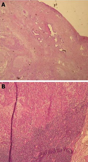
Figure 2 Histological examination.
A: Involvement of the cystic dilatation of common bile duct by the adenocarcinoma of gallbladder; B: Lymph node involvement by the adenocarcinoma of gallbladder.
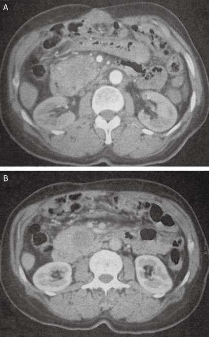
Figure 3 Abdominal CT: low-density mass in the pancreatic head.
A: Arterial phase; B: Portal phase. CT: Computed tomography.
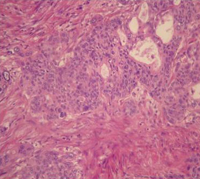
Figure 4 (He × 250): Microscopic findings: two components of the pancreatic carcinoma, the squamous one and the glandular one.
- Citation: Lahmar A, Abid SB, Arfa MN, Bayar R, Khalfallah MT, Mzabi-Regaya S. Metachronous cancer of gallbladder and pancreas with pancreatobiliary maljunction. World J Gastrointest Surg 2010; 2(4): 143-146
- URL: https://www.wjgnet.com/1948-9366/full/v2/i4/143.htm
- DOI: https://dx.doi.org/10.4240/wjgs.v2.i4.143


