Copyright
©The Author(s) 2025.
World J Gastrointest Surg. Aug 27, 2025; 17(8): 108579
Published online Aug 27, 2025. doi: 10.4240/wjgs.v17.i8.108579
Published online Aug 27, 2025. doi: 10.4240/wjgs.v17.i8.108579
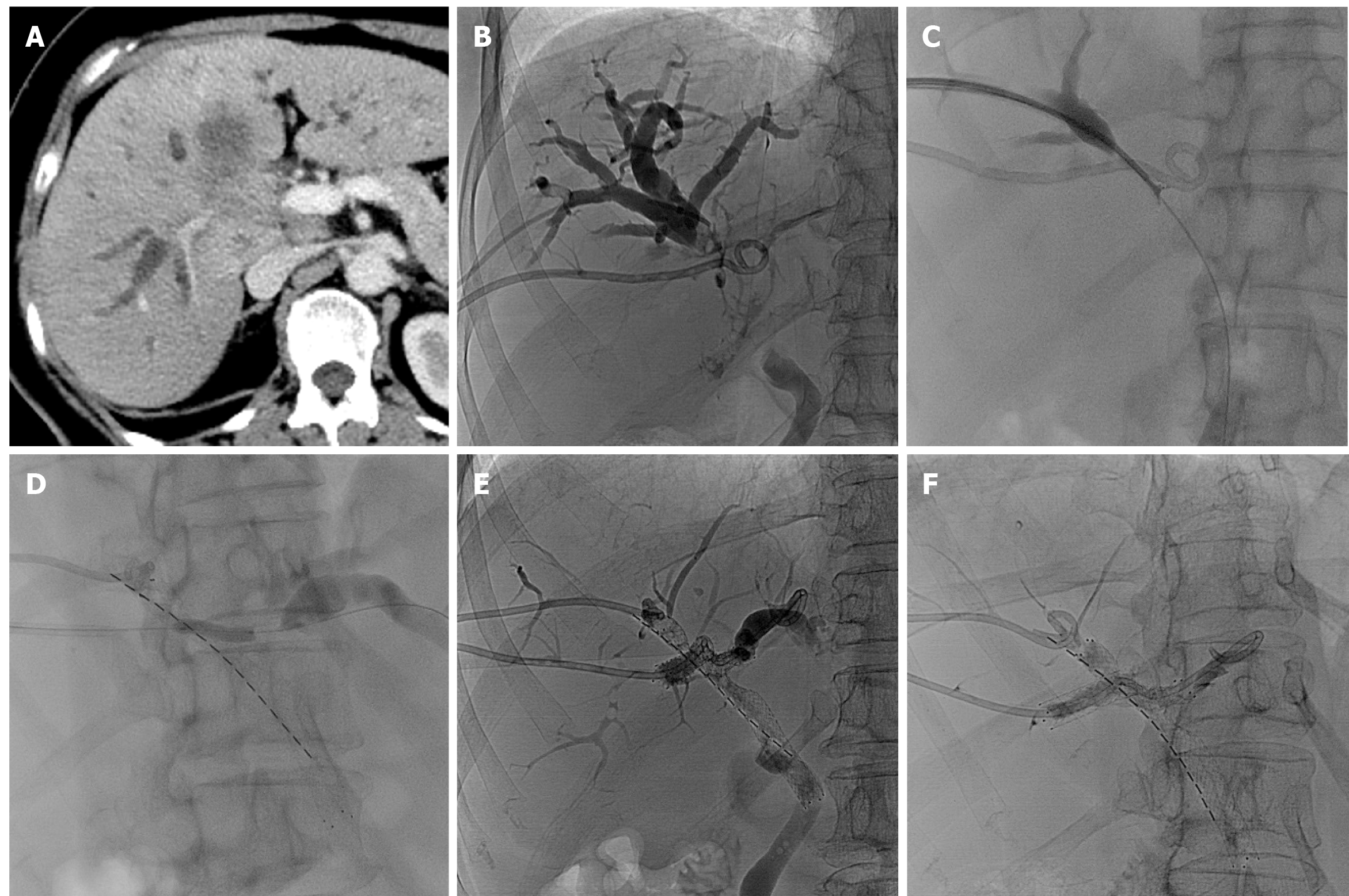
Figure 1 Gallbladder cancer invading the hepatic hilar in a 60-year-old female patient.
A: Abdominal contrast-enhanced computed tomography indicates gallbladder cancer invading the junction area of the left and right lobes of the liver, with intrahepatic bile duct dilation; B: Cholangiography shows hilar biliary obstruction, Bismuth type IV. Two biliary drainage tubes were inserted, with the tips of the drainage tubes located in the right anterior lobe bile duct and the left intrahepatic bile duct, respectively; C: Intraductal biopsy revealed adenocarcinoma; D: Double self-expandable metallic stent (SEMS) implantation (type X). The right anterior bile duct is connected to the common bile duct [8 mm × 80 mm, 16 iodine-125 (125I) seeds]. Subsequently, 4-mm and 6-mm balloons were used to dilate the severe stenosis segment (from the right posterior bile duct to the left bile duct). Implantation of an 8-mm × 60-mm SEMS was performed (from the right posterior bile duct to the left bile duct). Failure to implant 125I seed strips occurred due to severe tortuosity of the bile duct; E and F: Posterior-anterior/right anterior oblique 30-degree cholangiography indicates significant tortuosity from the right posterior bile duct to the left bile duct.
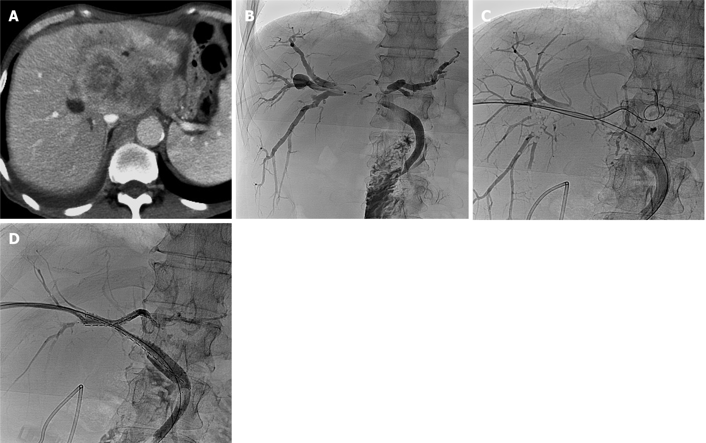
Figure 2 Liver metastasis and postoperative gastric cancer in a 61-years old male patient.
A: Abdominal contrast-enhanced computed tomography indicates an extensive malignant tumor in the hepatic left lobe, enlargement of the gallbladder, dilation of intrahepatic bile duct branches, abdominal and retroperitoneal lymph nodes metastasis, and ascites; B: Cholangiography shows hilar biliary obstruction, Bismuth type IV. Intraductal biopsy revealed adenocarcinoma; C: Double wires and double 6 French guiding tubes were selected for the common bile duct and the left intrahepatic bile duct, respectively; D: Double iodine-125 (125I) seed strip combined with double self-expandable metallic stent implantation (type T). The right bile duct is connected to the common bile duct (8 mm × 80 mm, 16 125I seeds); the right bile duct is connected to the left bile duct (8 mm × 60 mm, 14 125I seeds).
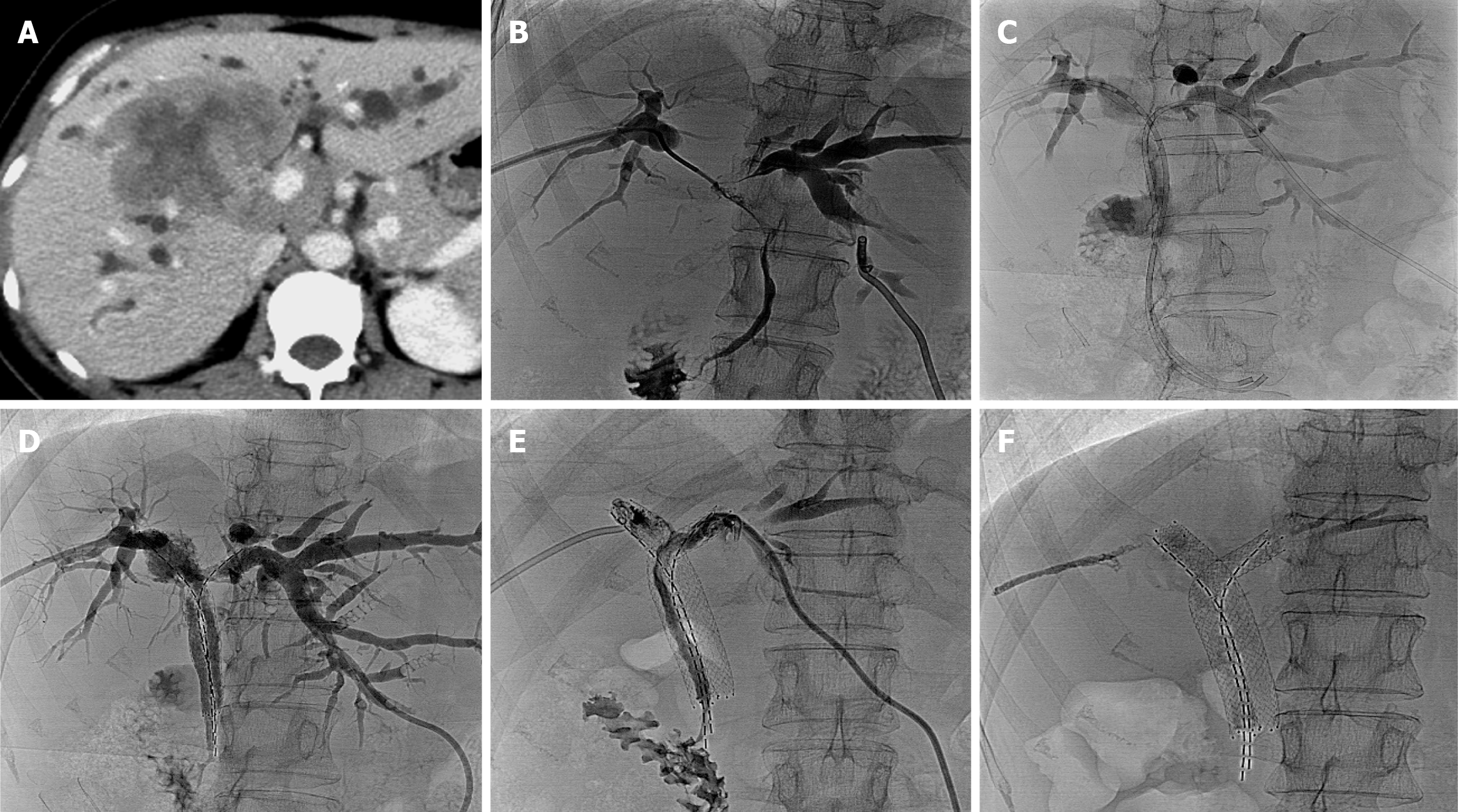
Figure 3 Hilar cholangiocarcinoma in a 49-year-old female patient.
A: Abdominal contrast-enhanced computed tomography shows hilar cho
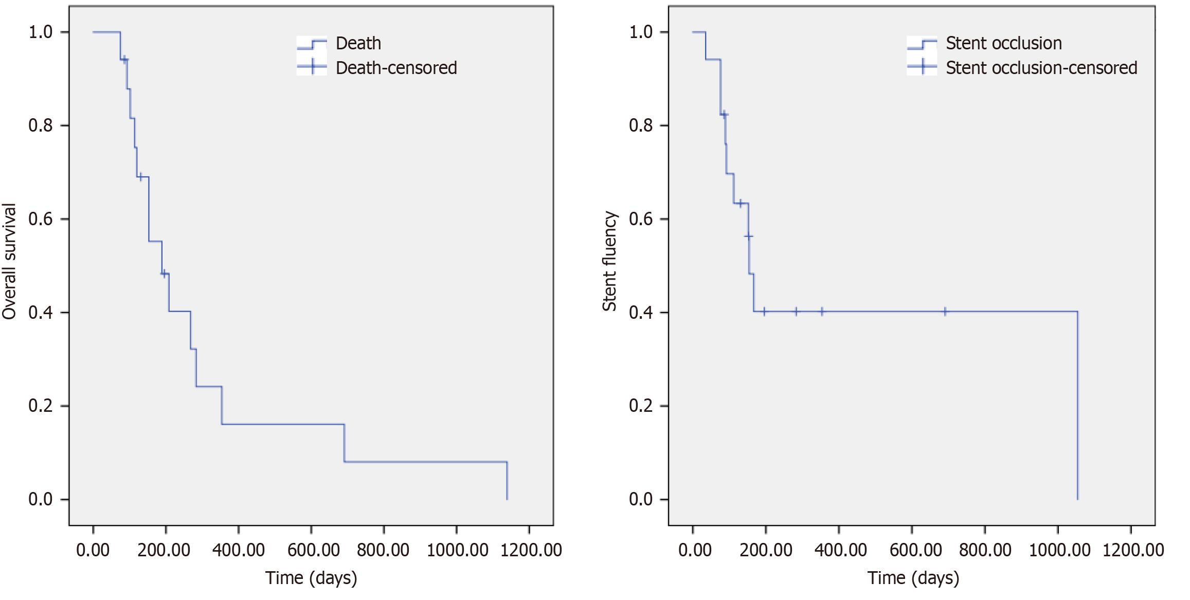
Figure 4
The overall survival was 189.
00 ± 47.27 days (95%CI: 96.35-281.66), and the stent fluency time was 154.00 ± 12.19 days (95%CI: 130.11-177.89).
- Citation: Zhou CG, Zhang Y, Li H, Liu KY, Yang XY, Gao K. Outcomes of iodine-125 seed strips combined with double self-expandable metallic stent for Bismuth type III and IV malignant biliary obstruction. World J Gastrointest Surg 2025; 17(8): 108579
- URL: https://www.wjgnet.com/1948-9366/full/v17/i8/108579.htm
- DOI: https://dx.doi.org/10.4240/wjgs.v17.i8.108579


