Copyright
©The Author(s) 2024.
World J Gastrointest Surg. Jun 27, 2024; 16(6): 1691-1699
Published online Jun 27, 2024. doi: 10.4240/wjgs.v16.i6.1691
Published online Jun 27, 2024. doi: 10.4240/wjgs.v16.i6.1691
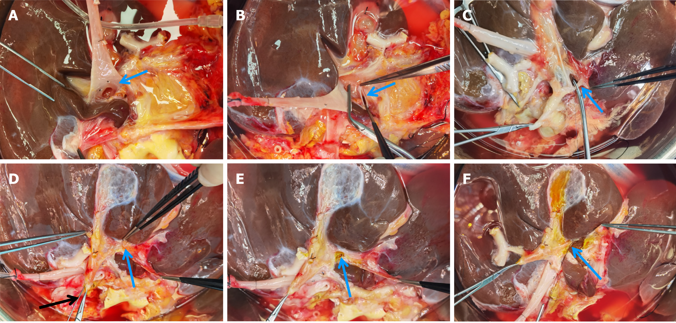
Figure 1 Procedure of splitting the first porta hepatis.
A: Separation of the left and right branches of the portal vein (arrow indicating the left portal vein); B: Division of the left portal vein (arrow) followed by suturing of the proximal end; C: Identification and division of the left hepatic artery (arrow); D: Identification of the division site of the left hepatic duct (blue arrow) under biliary probe guidance (black arrow); E: Incision of the left hepatic duct anterior wall (arrow) and reconfirmation of the left hepatic duct, right hepatic duct, and suspected bile duct openings using the probe; F: Division of the left hepatic duct, confirming the landmark for the division of liver parenchyma in the first porta hepatis.
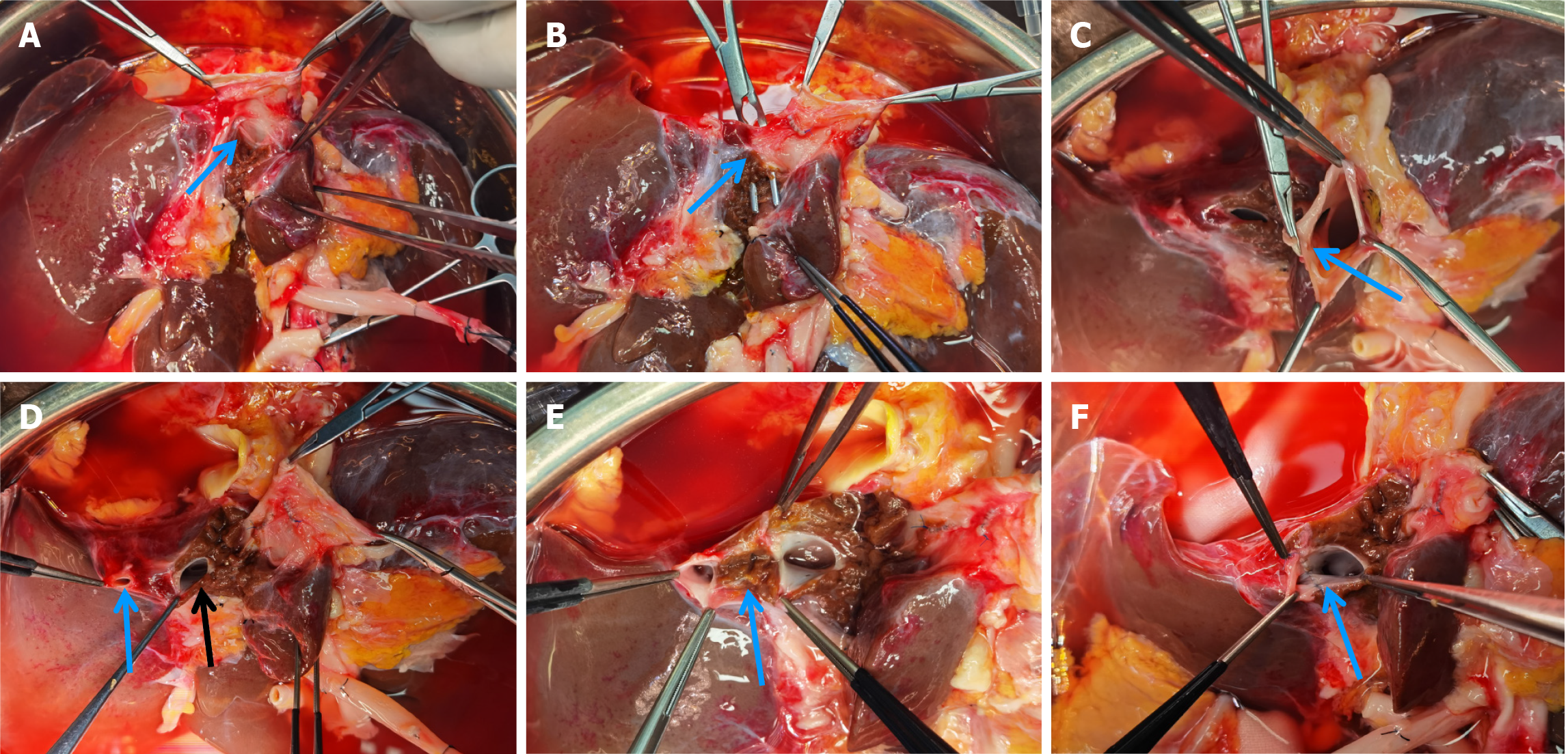
Figure 2 Step-by-step process involved in splitting the second porta hepatis.
A: Elevation of the suprahepatic inferior vena cava followed by blunt dissection of the liver tissue at the junction of the left hepatic vein and the inferior vena cava (arrow) to fully expose the left hepatic vein; B: Separating and dividing the left hepatic vein (arrow) using a vascular occlusion clamp on the inferior vena cava side; C: Formation of the middle hepatic vein and the opening of the inferior vena cava (arrow points to the formed vessel opening); D: Identification of the two openings of the left hepatic vein in the left lateral segment (blue arrow and black arrow); E: Removal of the liver tissue between the two openings of the left hepatic vein (arrow) to form a single opening; F: Display of the formed opening of the left hepatic vein.
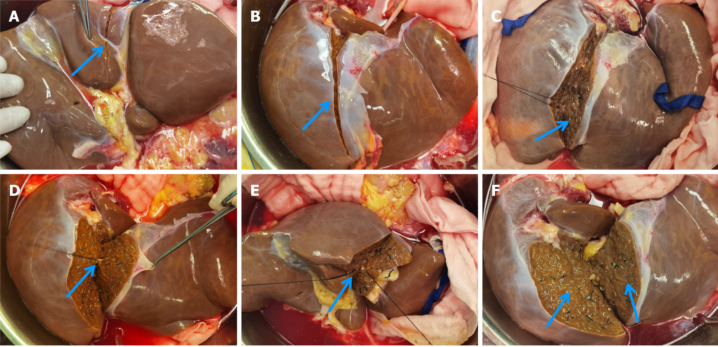
Figure 3 Procedure of liver parenchymal division.
A: Identification of the landmark line on the visceral surface of the liver for liver parenchymal division (0.5-1.0 cm to the right of the liver round ligament); B: Landmark line on the diaphragmatic surface of the liver for liver parenchymal division (on the right of the falciform ligament); C: Liver parenchymal division in the flat position, using titanium clips for small vessels (arrow); D: Adjusting the position of the liver during parenchymal division (suprahepatic inferior vena cava facing upwards), using silk sutures or ligatures for larger vessels (arrow); E: Continued liver parenchymal division with the liver flipped (suprahepatic inferior vena cava facing downward); F: Display of the two smooth liver segment surfaces after completion of the splitting process (arrow).
- Citation: Zhao D, Xie QH, Fang TS, Zhang KJ, Tang JX, Yan X, Jin X, Xie LJ, Xie WG. How to apply ex-vivo split liver transplantation safely and feasibly: A three-step approach. World J Gastrointest Surg 2024; 16(6): 1691-1699
- URL: https://www.wjgnet.com/1948-9366/full/v16/i6/1691.htm
- DOI: https://dx.doi.org/10.4240/wjgs.v16.i6.1691


