Copyright
©The Author(s) 2024.
World J Gastrointest Surg. Dec 27, 2024; 16(12): 3890-3894
Published online Dec 27, 2024. doi: 10.4240/wjgs.v16.i12.3890
Published online Dec 27, 2024. doi: 10.4240/wjgs.v16.i12.3890
Figure 1 Proximal transverse colon neoplasia.
Gross specimen following surgical resection of right-sided colon neoplasia after methylene blue and formaldehyde processing. A: Intraluminal aspect of the mucosa; B: Parasagittal serial slices with the localized lymph nodes in the mesocolon.
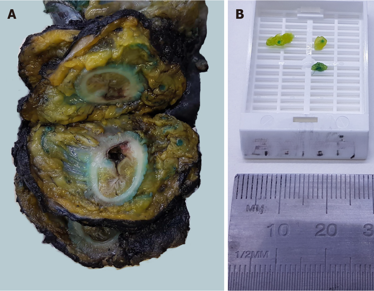
Figure 2 Rectum specimen and isolated lymph nodes.
Axial section of the specimen of rectal neoplasia. A: Stained lymph nodes are visualized in the mesorectum; B: Millimetric nodes are located within the fat tissue via blue methylene injection.
- Citation: Morera-Ocon FJ, Navarro-Campoy C, Cardona-Henao JD, Landete-Molina F. Colorectal cancer lymph node dissection and disease survival. World J Gastrointest Surg 2024; 16(12): 3890-3894
- URL: https://www.wjgnet.com/1948-9366/full/v16/i12/3890.htm
- DOI: https://dx.doi.org/10.4240/wjgs.v16.i12.3890


