Copyright
©The Author(s) 2024.
World J Gastrointest Surg. Dec 27, 2024; 16(12): 3870-3874
Published online Dec 27, 2024. doi: 10.4240/wjgs.v16.i12.3870
Published online Dec 27, 2024. doi: 10.4240/wjgs.v16.i12.3870
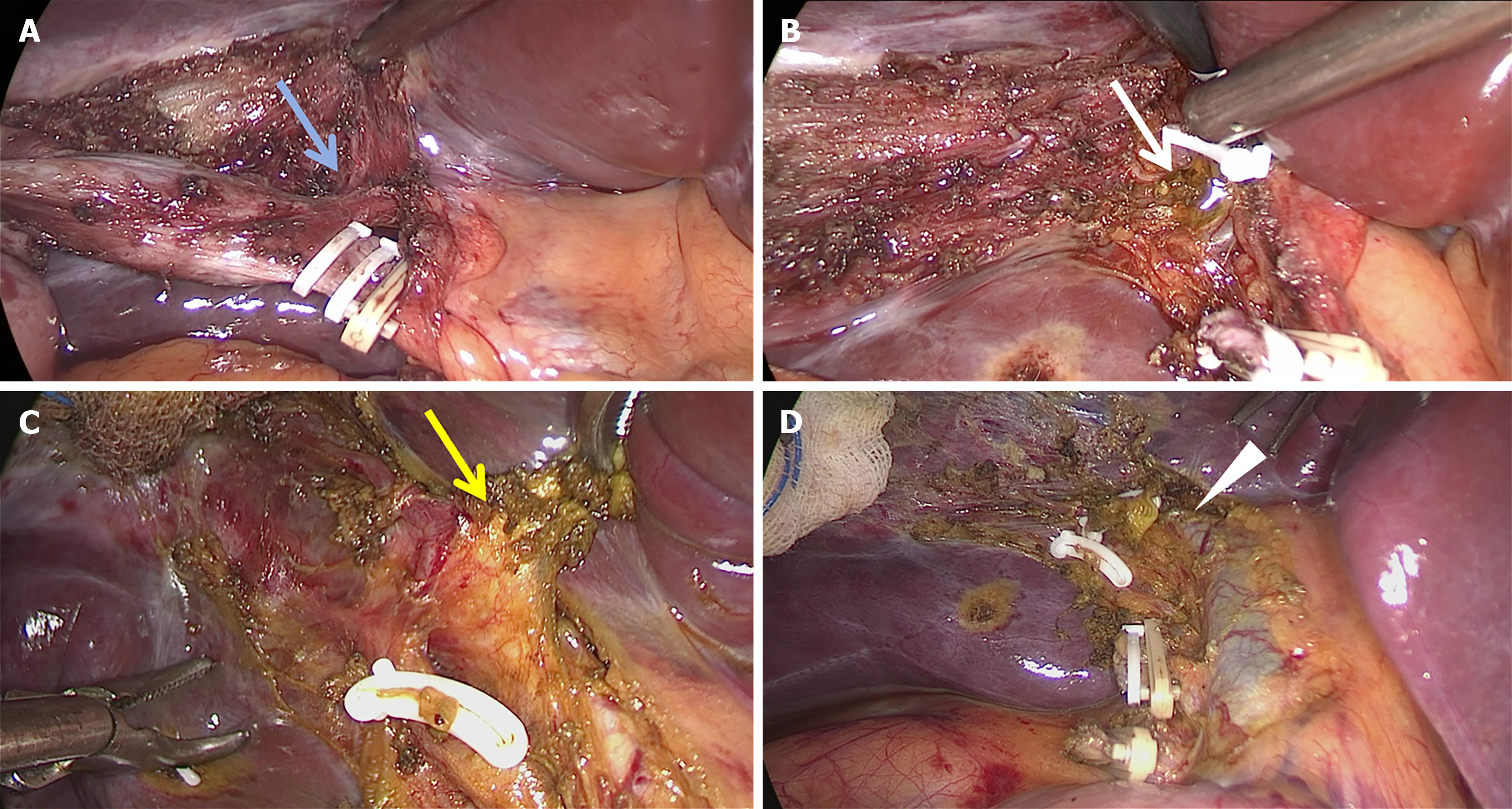
Figure 1 Laparoscopic view during cholecystectomy.
A: Anomalous duct (blue arrow) was found extending from the gallbladder ampulla to inside the liver; B: Bile leak (white arrow) was detected from the cut end of the anomalous duct, confirming it is an aberrant bile duct; C and D: Confirmation that the other end (yellow arrow) of the injured bile duct was connected to the right hepatic duct (white arrowhead), identifying it as a communication accessory hepatic duct, which was clipped with a vascular clamp.
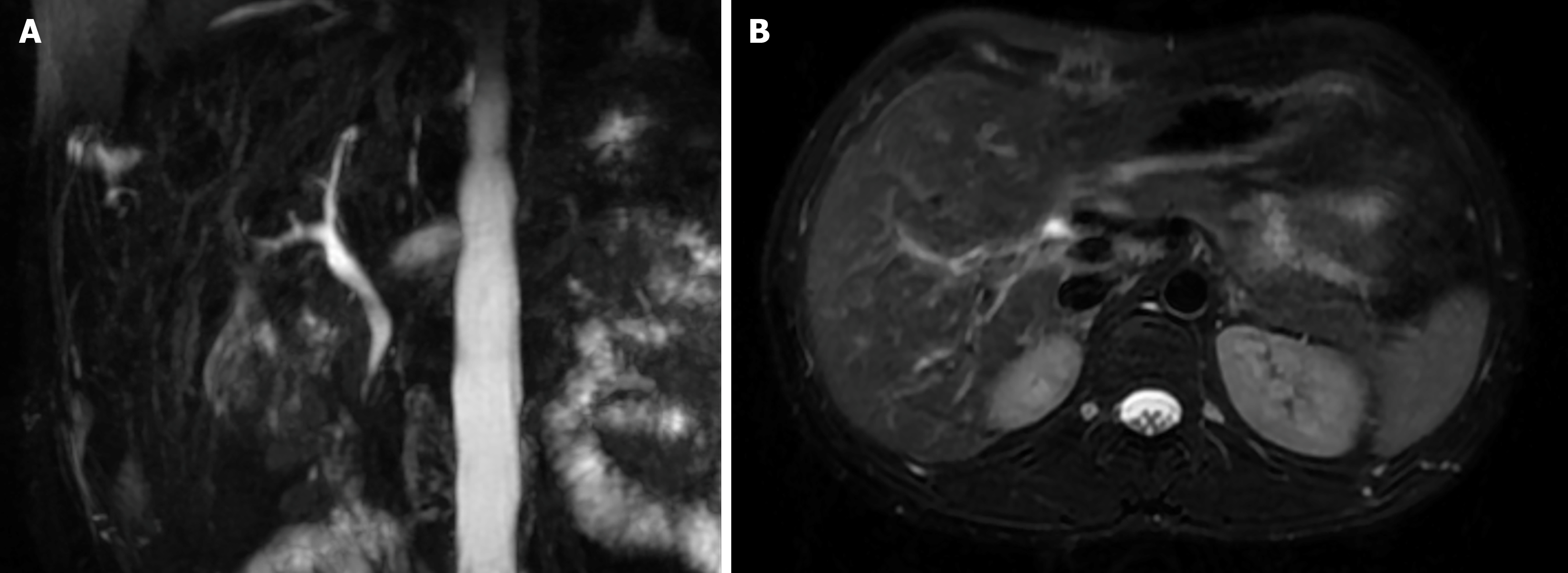
Figure 2 Magnetic resonance imaging and magnetic resonance cholangiopancreatography after surgery.
A and B: Two weeks after surgery, follow-up magnetic resonance imaging and magnetic resonance cholangiopancreatography showed no intrahepatic or extrahepatic bile duct strictures or dilatation.
- Citation: Zhao PJ, Ma Y, Yang JW. Laparoscopic cholecystectomy with communicating accessory hepatic duct injury and management: A case report. World J Gastrointest Surg 2024; 16(12): 3870-3874
- URL: https://www.wjgnet.com/1948-9366/full/v16/i12/3870.htm
- DOI: https://dx.doi.org/10.4240/wjgs.v16.i12.3870


