Copyright
©The Author(s) 2024.
World J Gastrointest Surg. Dec 27, 2024; 16(12): 3862-3869
Published online Dec 27, 2024. doi: 10.4240/wjgs.v16.i12.3862
Published online Dec 27, 2024. doi: 10.4240/wjgs.v16.i12.3862
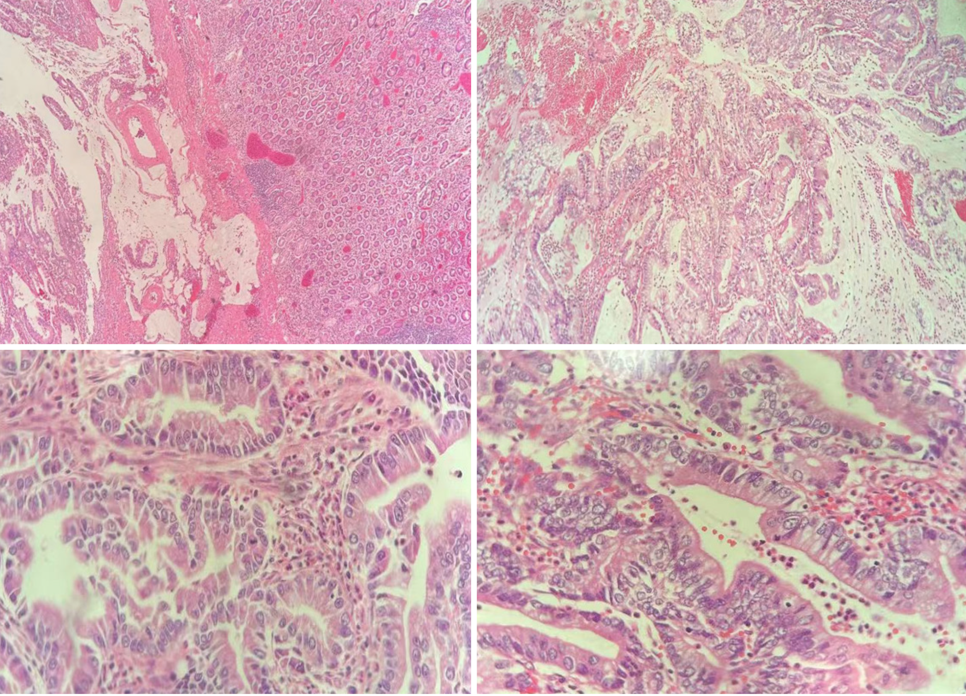
Figure 1 Pathological features of descending duodenal mucosal adenocarcinoma.
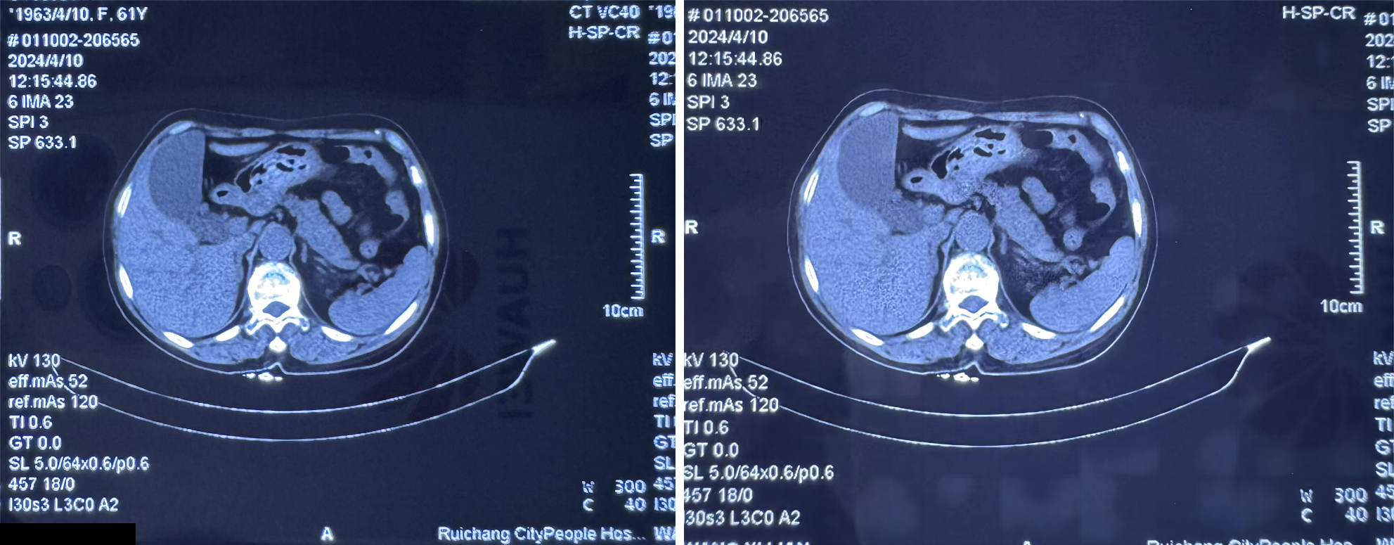
Figure 2 Results of plain computed tomography scan of the patient’s lower abdomen.
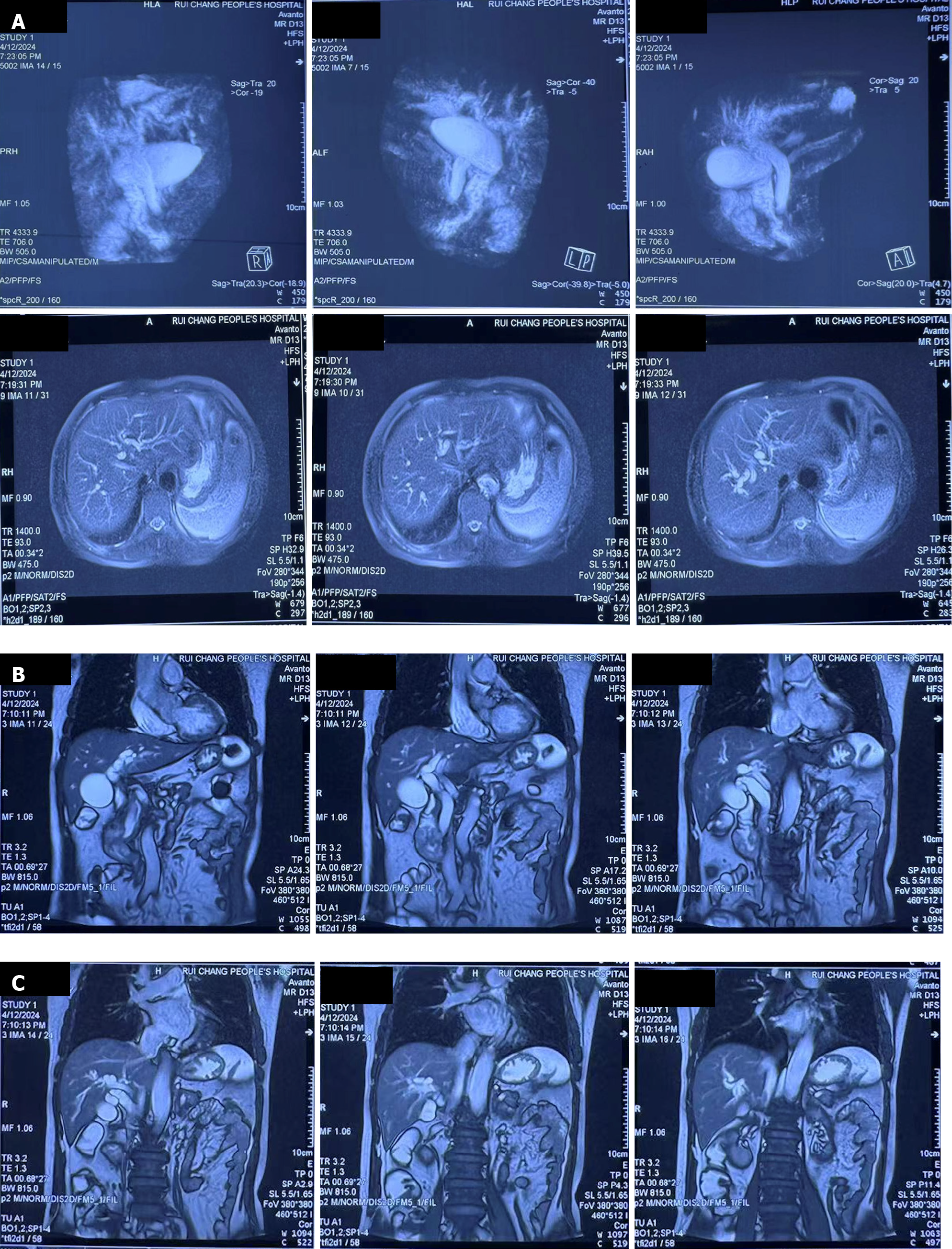
Figure 3 Magnetic resonance imaging images of the patient’s lower abdomen.
A: Cross section and side view; B: Anterior coronal plane; C: Posterior coronal plane.
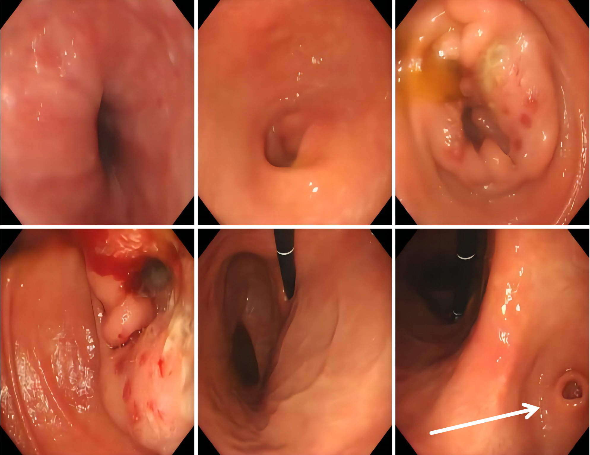
Figure 4 Electronic proctoscopy image results.
Arrow: Lesion of coronal plane.
- Citation: Zhang JY, Wu LS, Yan J, Jiang Q, Li XQ. Pathological diagnosis and clinical feature analysis of descending duodenal mucosal adenocarcinoma: A case report. World J Gastrointest Surg 2024; 16(12): 3862-3869
- URL: https://www.wjgnet.com/1948-9366/full/v16/i12/3862.htm
- DOI: https://dx.doi.org/10.4240/wjgs.v16.i12.3862


