Copyright
©The Author(s) 2024.
World J Gastrointest Surg. Dec 27, 2024; 16(12): 3806-3817
Published online Dec 27, 2024. doi: 10.4240/wjgs.v16.i12.3806
Published online Dec 27, 2024. doi: 10.4240/wjgs.v16.i12.3806
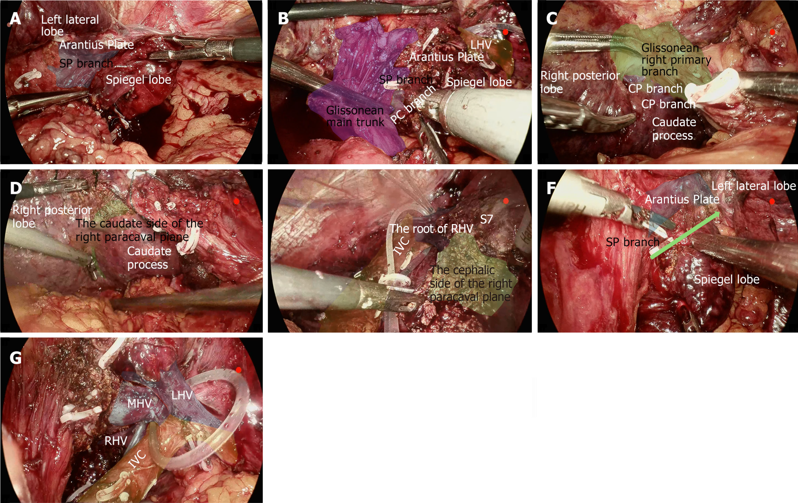
Figure 1 Segmentectomy 1.
A: Dissecting the spiegel lobe branch; B: Dissecting the paracaval portion branch; C: Dissecting the caudate process branch; D: Liver parenchyma resection along the markings; E: Liver parenchyma resection along the markings; F: Liver parenchyma resection along the markings; G: Liver parenchyma resection along the markings. SP: Spiegel lobe; CP: Caudate process; LHV: Left hepatic vein; MHV: Middle hepatic vein; RHV: Right hepatic vein; IVC: Inferior vena cava; S7: Segment 7.
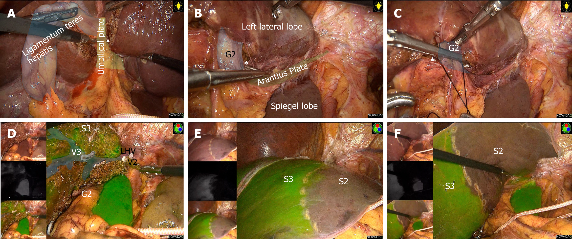
Figure 2 Segmentectomy 2.
A: Beginning from the umbilical plate; B: Exposing the caudal end of the Arantius plate; C: Dissecting the Glissonean pedicle of segment 2; D: Liver parenchyma resection along the markings; E and F: Indocyanine green staining. G2: Glissonean pedicle of segment 2; S2: Segment 2; S3: Segment 3; V3: Hepatic vein of segment 3; LHV: Left hepatic vein.
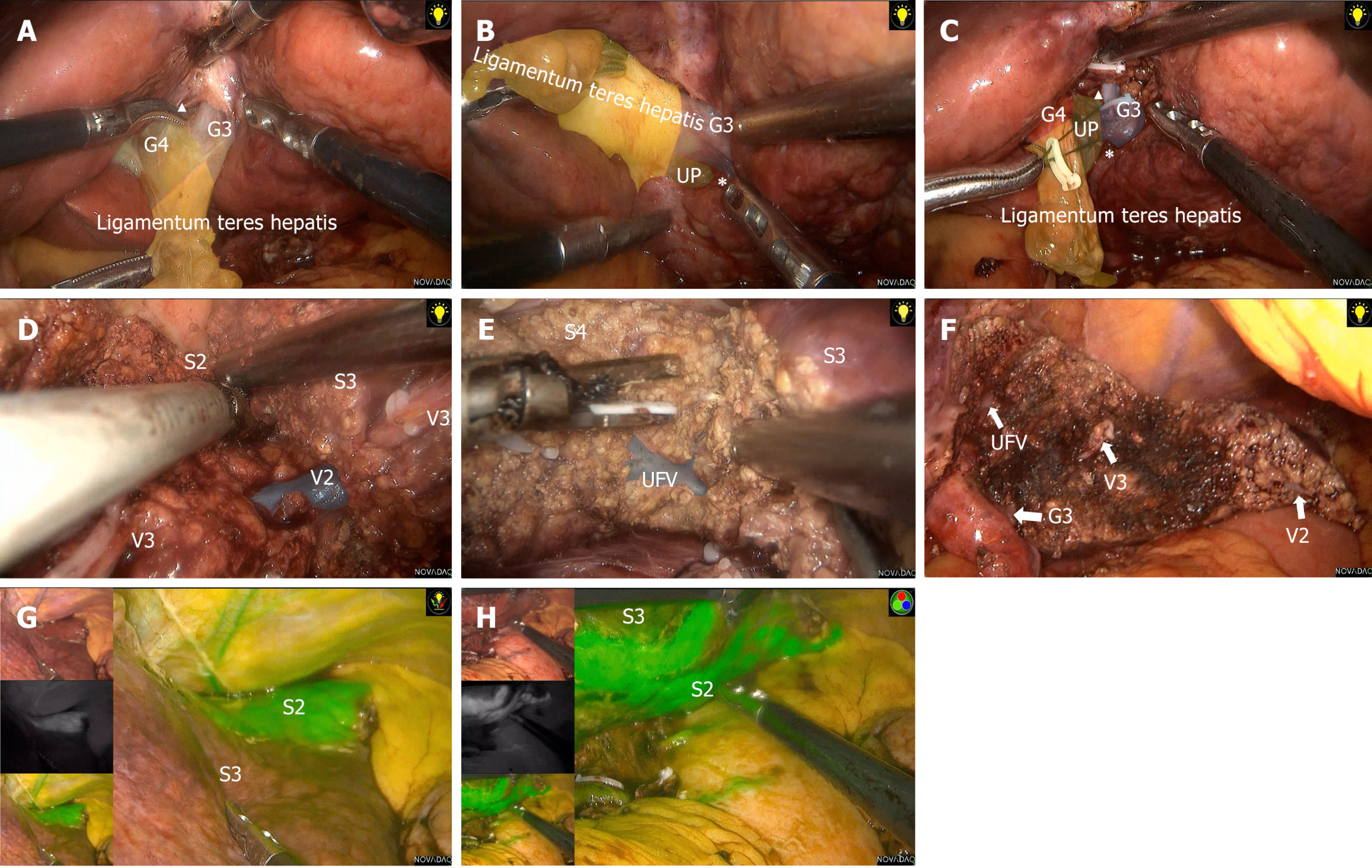
Figure 3 Segmentectomy 3.
A: Exposing the root of the round ligament; B: Exposing the umbilical plate; C: Dissecting the Glissonean pedicle of segment 3; D: Liver parenchyma resection along the markings; E: Liver parenchyma resection along the markings; F: Segment 3 resection is completed; G and H: Indocyanine green staining. G3: Glissonean pedicle of segment 3; G4: Glissonean pedicle of segment 4; V2: Hepatic vein of segment 2; UFV: Umbilical fissure vein; UP: Umbilical plate; S3: Segment 3.
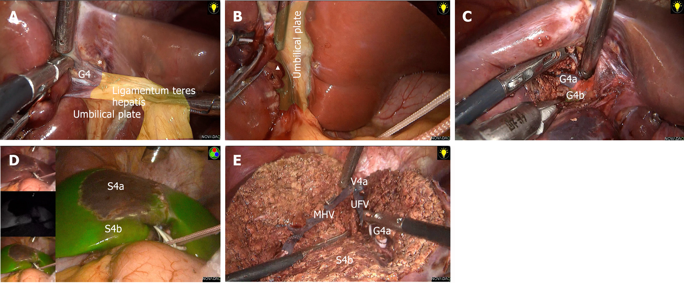
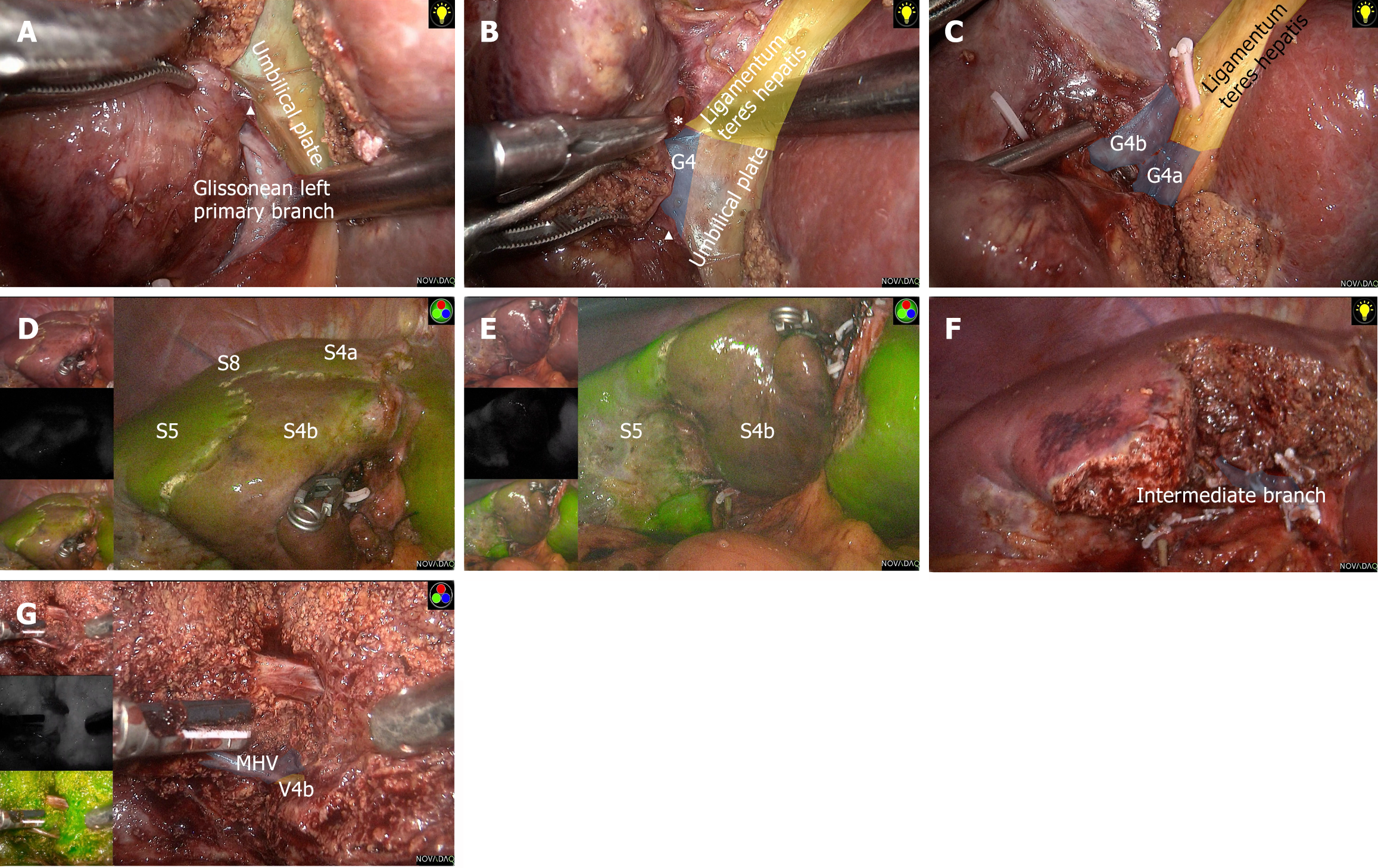
Figure 4 Subsegmentectomy 4a.
A: Exposing the root of the round ligament; B: Exposing the umbilical plate; C: Dissecting the Glissonean pedicle of segment 4a; D: Indocyanine green staining; E: Segment 4a resection is completed. G4: Glissonean pedicle of segment 4; G4b: Glissonean pedicle of segment 4b; G4a: Glissonean pedicle of segment 4a; MHV: Middle hepatic vein; UFV: Umbilical fissure vein; S4a: Segment 4a; S4b: Segment 4b; V4a: Hepatic vein of segment 4a.
Figure 5 Subsegmentectomy 4b.
A: Exposing the umbilical plate; B: Exposing the root of the round ligament; C: Dissecting the Glissonean pedicle of segment 4b; D and E: Indocyanine green staining; F: The intermediate branch; G: Segment 4b resection is completed. G4: Glissonean pedicle of segment 4; G4b: Glissonean pedicle of segment 4b; G4a: Glissonean pedicle of segment 4a; MHV: Middle hepatic vein; S5: Segment 5; S8: Segment 8; V4b: Hepatic vein of segment 4b.
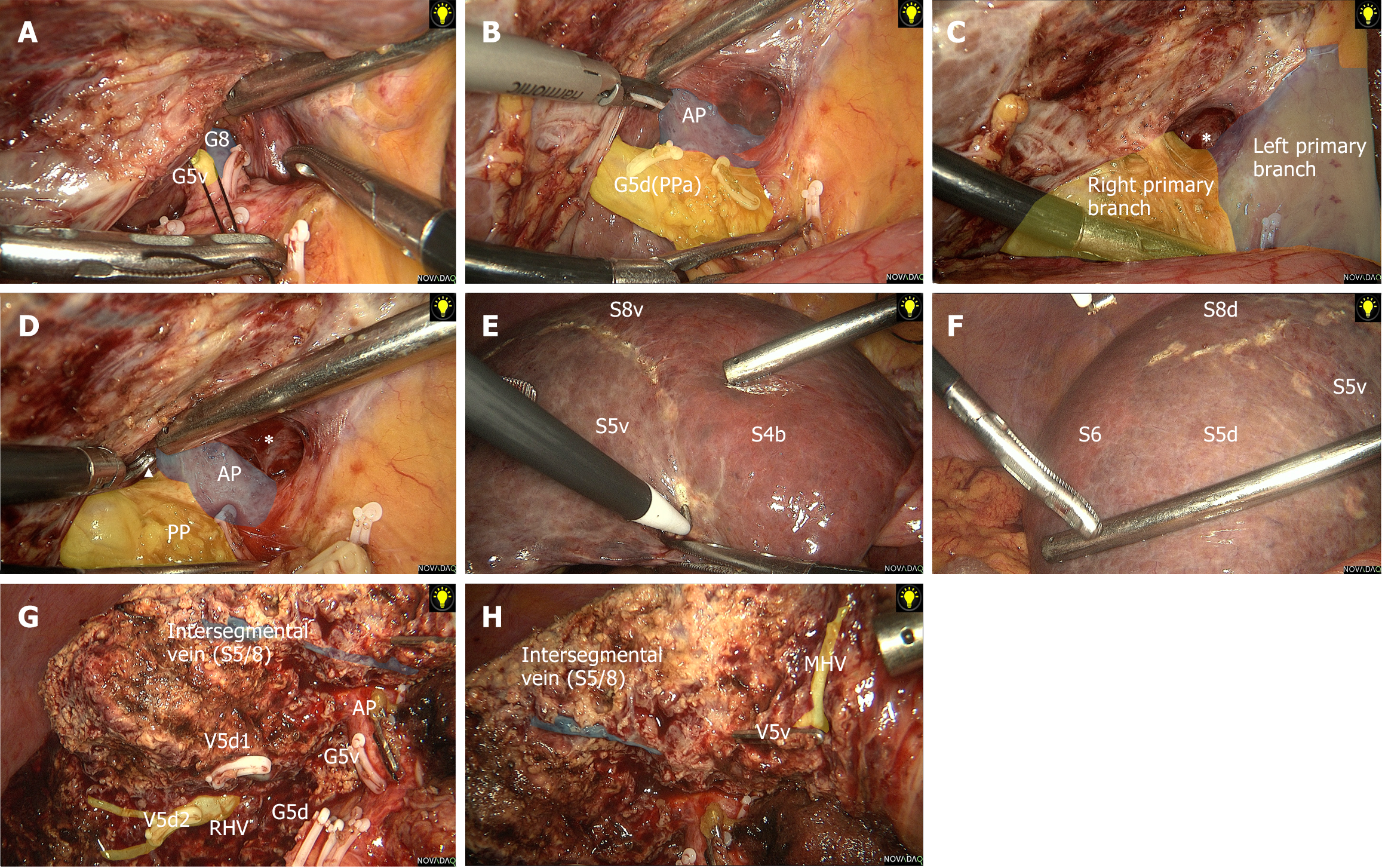
Figure 6 Segmentectomy 5.
A: Dissecting the Glissonean pedicle of segment 5 ventral; B: Dissecting the Glissonean pedicle of segment 5 dorsal; C and D: Lowering the hepatic hilar plate; E: The ischemic line (between segment 5 ventral portion and segment 4b); F: The ischemic line (between segment 5d and segment 6); G: Segment 5 resection is completed; H: Segment 5 resection is completed. AP: Anterior pedicle; PP: Posterior pedicle; G8: Glissonean pedicle of segment 8; G5v: Glissonean pedicle of segment 5 ventral; G5d: Glissonean pedicle of segment 5 dorsal; RHV: Right hepatic vein; MHV: Middle hepatic vein; V5v: Hepatic vein of segment 5 ventral portion; V5d: Hepatic vein of segment 5 dorsal portion; S5v: Segment 5 ventral portion; S5d: Segment 5 dorsal portion; S6: Segment 6; S8v: Segment 8 ventral portion; S8d: Segment 8 dorsal portion; PPa: First caudal lateral branch of the right posterior pedicle.
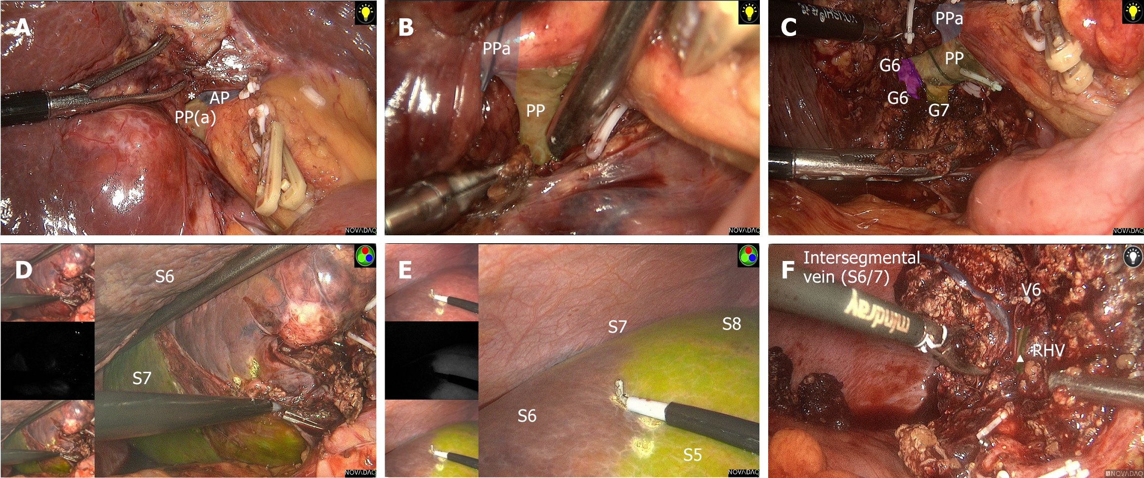
Figure 7 Segmentectomy 6.
A and B: Lowering the hepatic hilar plate; C: Dissecting the Glissonean pedicle of segment 6; D and E: Indocyanine green staining; F: Segment 6 resection is completed. AP: Anterior pedicle; PP: Posterior pedicle; RHV: Right hepatic vein; G6: Glissonean pedicle of segment 6; S6: Segment 6.
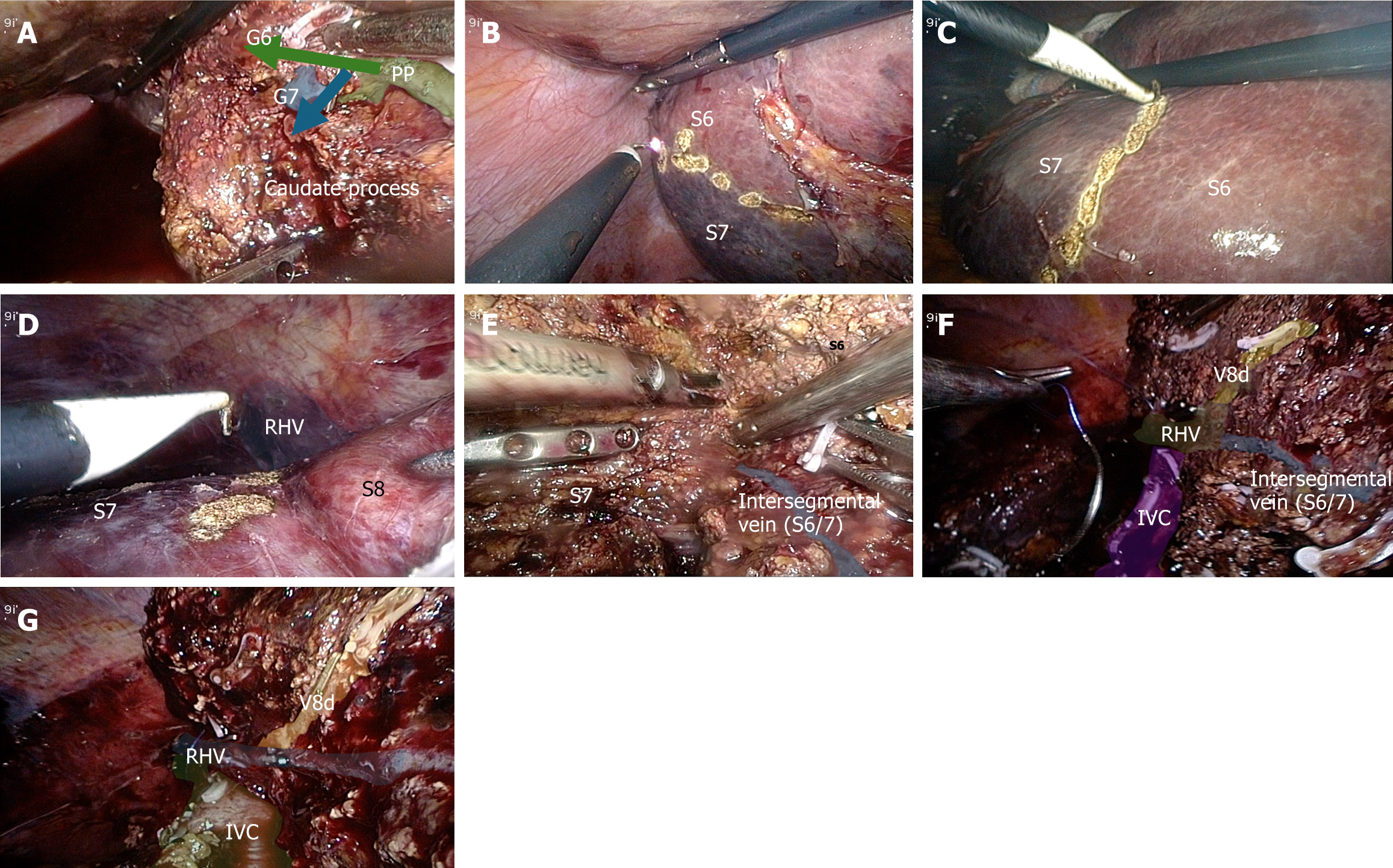
Figure 8 Segmentectomy 7.
A: Dissecting the Glissonean pedicle of segment 7; B: The ischemic line (dorsal side between segment 6 and segment 7); C: The ischemic line (ventral side between segment 6 and segment 7); D: The ischemic line (between segment 7 and segment 8); E: Liver parenchyma resection along the markings; F and G: Segment 7 resection is completed. V8d: Hepatic vein of segment 8 dorsal portion. G6: Glissonean pedicle of segment 6; PP: Posterior pedicle; RHV: Right hepatic vein; IVC: Inferior vena cava; S6: Segment 6.
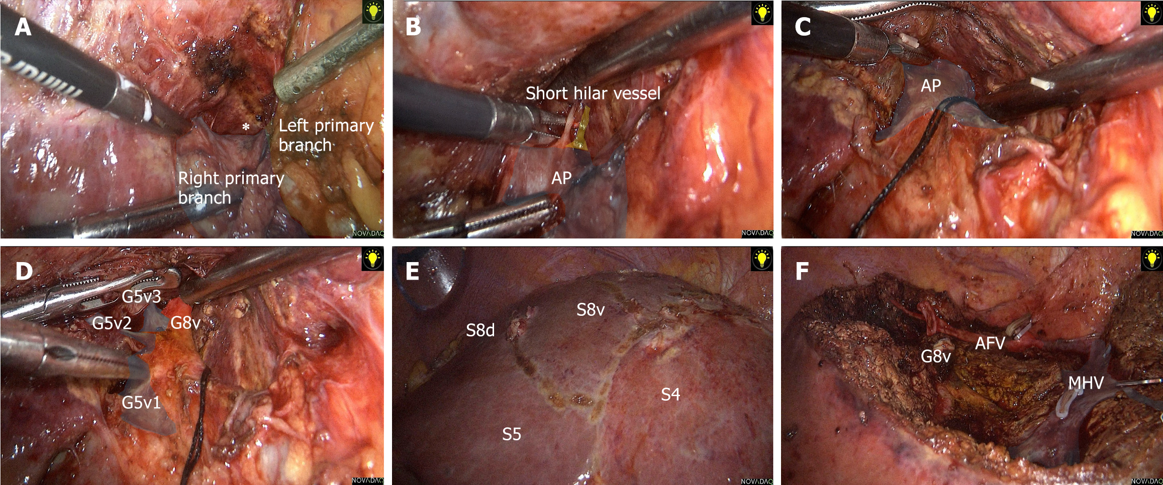
Figure 9 Subsegmentectomy 8 ventral portion.
A-C: Lowering the hepatic hilar plate (A and C), the short hilar vessel is transected (B); D: Dissecting the Glissonean pedicle of segment 8 ventral portion; E: The ischemic line; F: Segment 8 ventral resection is completed. AP: anterior pedicle; G8v: Glissonean pedicle of segment 8 ventral portion; S4: Segment 4; AFV: Anterior fissure vein; S8d: Segment 8 dorsal; S8v: Segment 8 ventral; MHV: Middle hepatic vein.
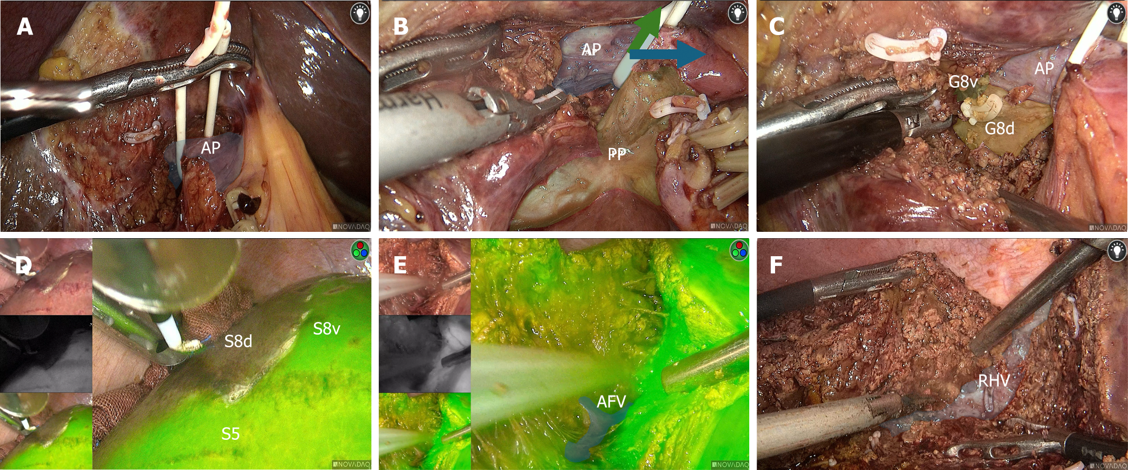
Figure 10 Subsegmentectomy 8 dorsal portion.
A: Dissecting the right anterior pedicle; B: Pulling the right anterior pedicle; C: Dissecting the Glissonean pedicle of segment 8 dorsal; D: Indocyanine green staining; E: Liver parenchyma resection along the markings; F: Segment 8 dorsal resection is completed. AP: Anterior pedicle; PP: Posterior pedicle; G8d: Glissonean pedicle of segment 8 dorsal; G8v: Glissonean pedicle of segment 8 ventral; S5: Segment 1; S8d: Segment 8 dorsal; S8v: Segment 8 ventral; AFV: Anterior fissure vein; RHV: Right hepatic vein.
- Citation: Wang SD, Wang L, Xiao H, Chen K, Liu JR, Chen Z, Lan X. Novel techniques of liver segmental and subsegmental pedicle anatomy from segment 1 to segment 8. World J Gastrointest Surg 2024; 16(12): 3806-3817
- URL: https://www.wjgnet.com/1948-9366/full/v16/i12/3806.htm
- DOI: https://dx.doi.org/10.4240/wjgs.v16.i12.3806


