Copyright
©The Author(s) 2024.
World J Gastrointest Surg. Dec 27, 2024; 16(12): 3780-3785
Published online Dec 27, 2024. doi: 10.4240/wjgs.v16.i12.3780
Published online Dec 27, 2024. doi: 10.4240/wjgs.v16.i12.3780
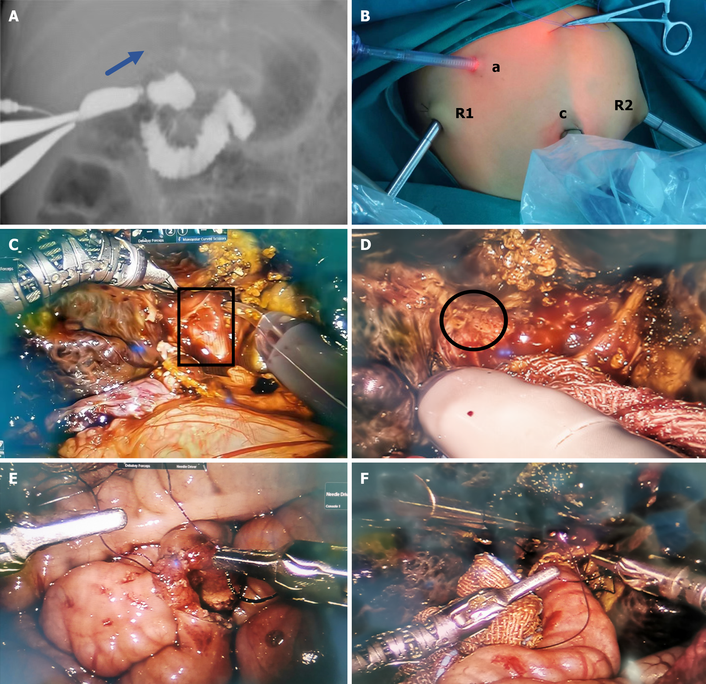
Figure 1 Intraoperative pictures.
A: Laparoscopic-assisted cholangiography indicated non-visualization of the intrahepatic and hepatic portal bile ducts; B: The camera port (c, 8.5 mm), No. 1 operation port (R1, 8 mm), No. 2 operation port (R2, 8 mm), accessory port (a, 5 mm); C: Fibrous cone at the hepatic porta (black box); D: Micro-bile ducts (black circle); E: End-to-side anastomosis in the abdominal cavity; F: Anastomosis between porta hepatis and jejunum.
- Citation: Xing GD, Wang XQ, Duan L, Liu G, Wang Z, Xiao YH, Xia Q, Xie HW, Shen Z, Yu ZZ, Huang LM. Robotic-assisted Kasai portoenterostomy for child biliary atresia. World J Gastrointest Surg 2024; 16(12): 3780-3785
- URL: https://www.wjgnet.com/1948-9366/full/v16/i12/3780.htm
- DOI: https://dx.doi.org/10.4240/wjgs.v16.i12.3780


