Copyright
©The Author(s) 2024.
World J Gastrointest Surg. Dec 27, 2024; 16(12): 3703-3709
Published online Dec 27, 2024. doi: 10.4240/wjgs.v16.i12.3703
Published online Dec 27, 2024. doi: 10.4240/wjgs.v16.i12.3703
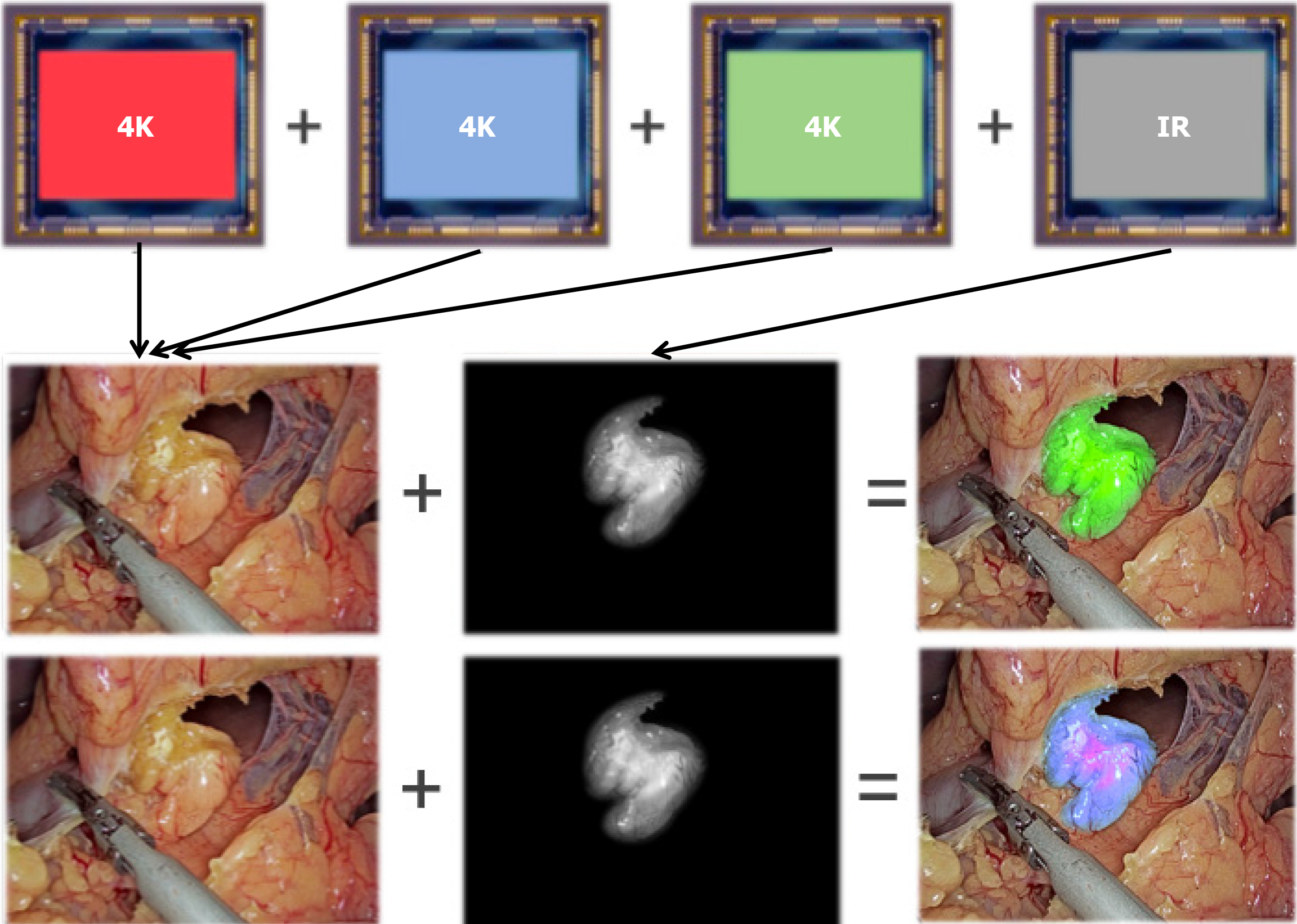
Figure 1 Principles and visual representation of multi-color fluorescence imaging.
The fluorescence intensity signal of bile ducts and the liver was represented by different colors among the blue-purple range. It was compared to the traditional laparoscopy images with single-color fluorescence.
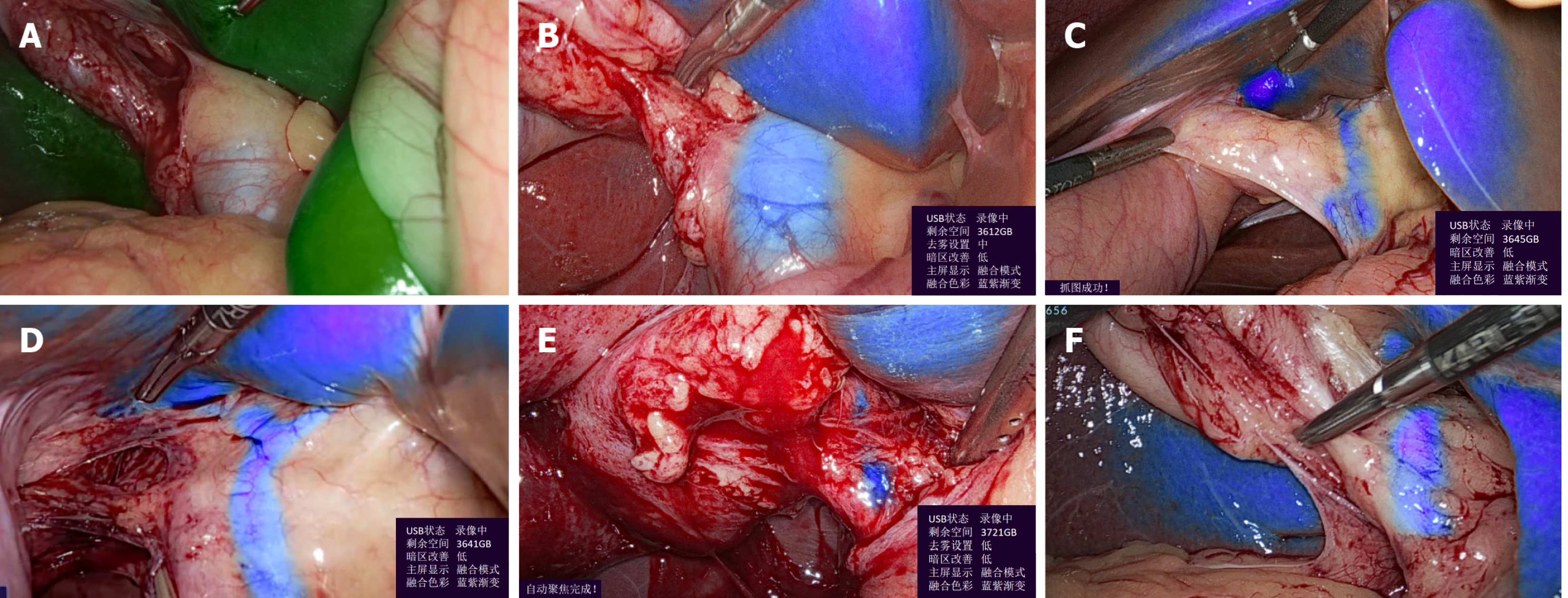
Figure 2 Clinical scenes of multi-color gradient fluorescence imaging.
A: Demonstrated the situation where the single color fluorescence imaging (SCFI) could not show the common bile duct; B: The situation where the SCFI could not show the common bile duct could be identified with the help of multi-color fluorescence imaging (MCFI); C: Showed the shape of the left and right hepatic ducts hidden in the liver tissue assisted by MCFI; D: Another setting showing the shape of the left and right hepatic ducts hidden in the liver tissue assisted by MCFI; E: Showed the structure of liver and the common bile duct in the situation of tissue adhesion and bleeding; F: MCFI would not be disturbed by the shades of surgical instruments and could show the structure of the common bile duct.
- Citation: Li JY, Ping L, Lin BZ, Wang ZH, Fang CH, Hua SR, Han XL. Efficacy of multi-color near-infrared fluorescence with indocyanine green: A new imaging strategy and its early experience in laparoscopic cholecystectomy. World J Gastrointest Surg 2024; 16(12): 3703-3709
- URL: https://www.wjgnet.com/1948-9366/full/v16/i12/3703.htm
- DOI: https://dx.doi.org/10.4240/wjgs.v16.i12.3703


