Copyright
©The Author(s) 2024.
World J Gastrointest Surg. Dec 27, 2024; 16(12): 3685-3693
Published online Dec 27, 2024. doi: 10.4240/wjgs.v16.i12.3685
Published online Dec 27, 2024. doi: 10.4240/wjgs.v16.i12.3685
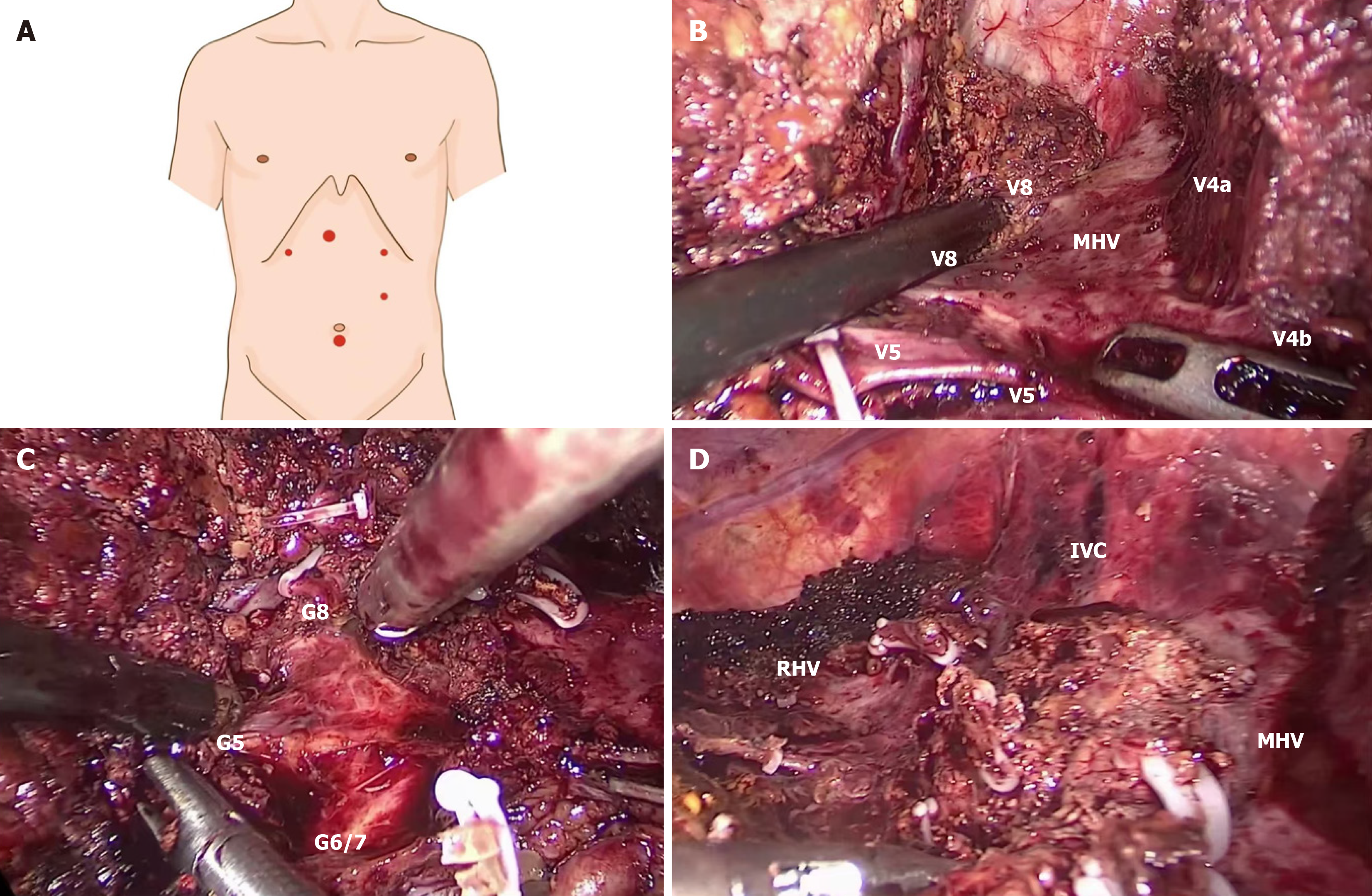
Figure 1 SVIII resection from caudal side.
A: Port placement for SVIII resection from caudal side; B: Dissection the middle hepatic vein from caudal side; C: Exposing G8 from caudal side; D: Photograph after SVIII resection from caudal side. MHV: Middle hepatic vein; RHV: Right hepatic vein.
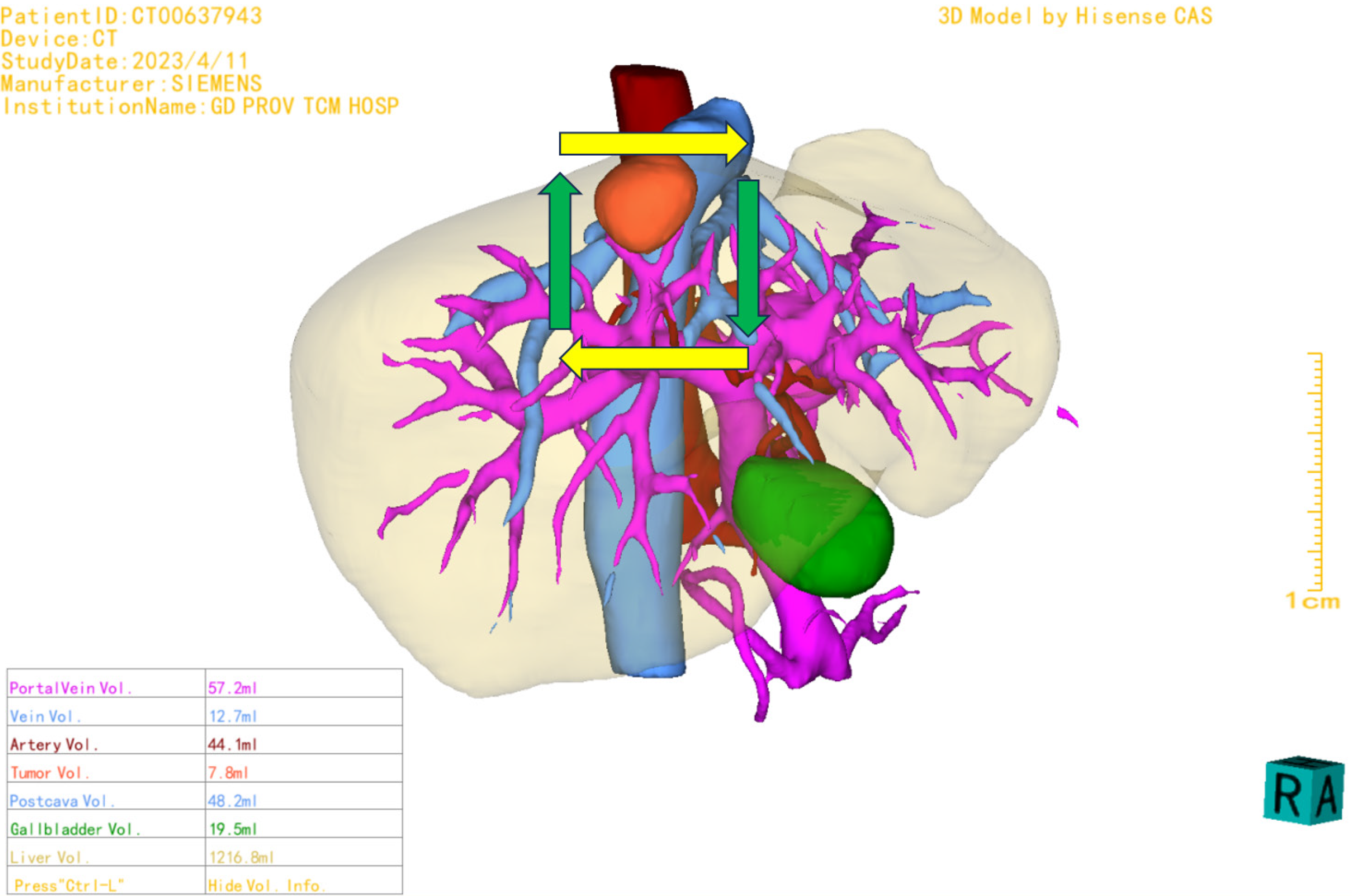
Figure 2 Three-dimensional reconstruction before the surgery.
3D: Three-dimensional.
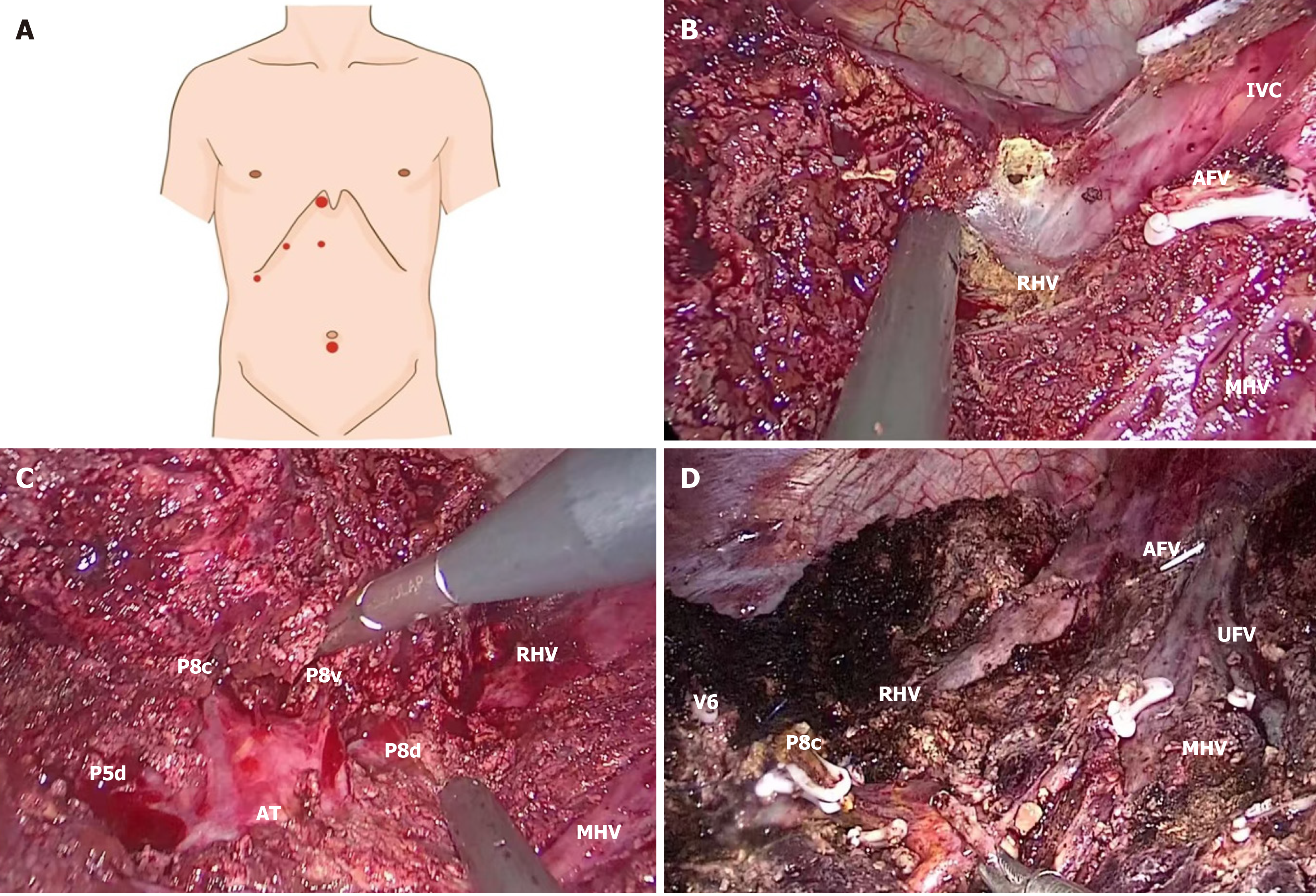
Figure 3 SVIII resection from cranio side.
A: Port placement for SVIII resection from cranio side; B: Dissection the middle hepatic vein from cranio side; C: Exposing G8 from cranio side; D: Photograph after SVIII resection from cranio side. MHV: Middle hepatic vein; RHV: Right hepatic vein; AFV: Anterior fissure vein; IVC: Inferior vena cava; UFV: Umbilical fissure vein.
- Citation: Peng JX, Li HL, Ye Q, Mo JQ, Wang JY, Liu ZY, He JM. Laparoscopic anatomical SVIII resection via middle hepatic fissure approach: Caudal or cranio side. World J Gastrointest Surg 2024; 16(12): 3685-3693
- URL: https://www.wjgnet.com/1948-9366/full/v16/i12/3685.htm
- DOI: https://dx.doi.org/10.4240/wjgs.v16.i12.3685


