Copyright
©The Author(s) 2024.
World J Gastrointest Surg. Oct 27, 2024; 16(10): 3301-3311
Published online Oct 27, 2024. doi: 10.4240/wjgs.v16.i10.3301
Published online Oct 27, 2024. doi: 10.4240/wjgs.v16.i10.3301
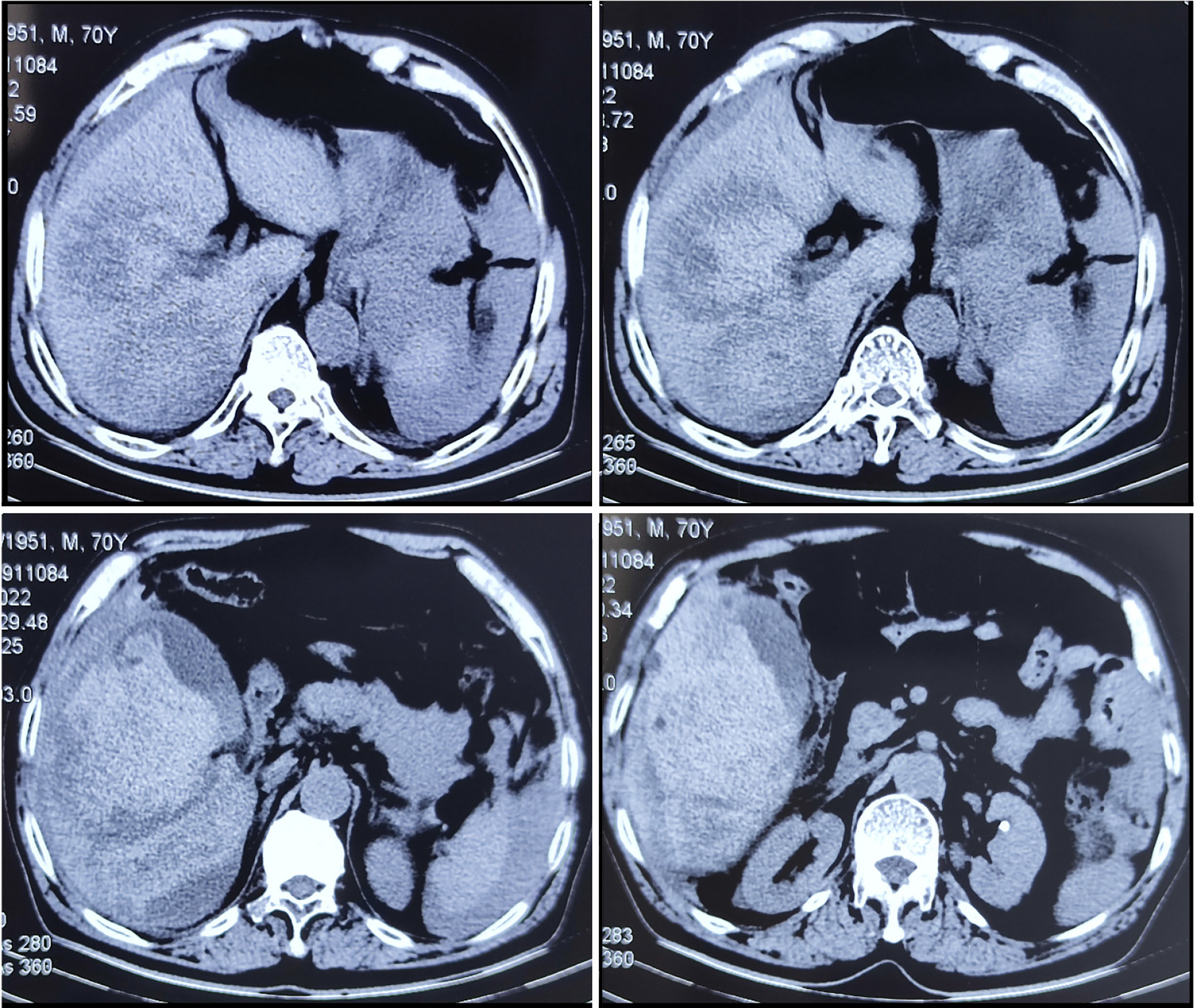
Figure 1 Abdominal computed tomography revealing a mixed density shadow in the right lobe of the liver combined with abdominopelvic effusion and an enlarged gallbladder adjoining the mass.
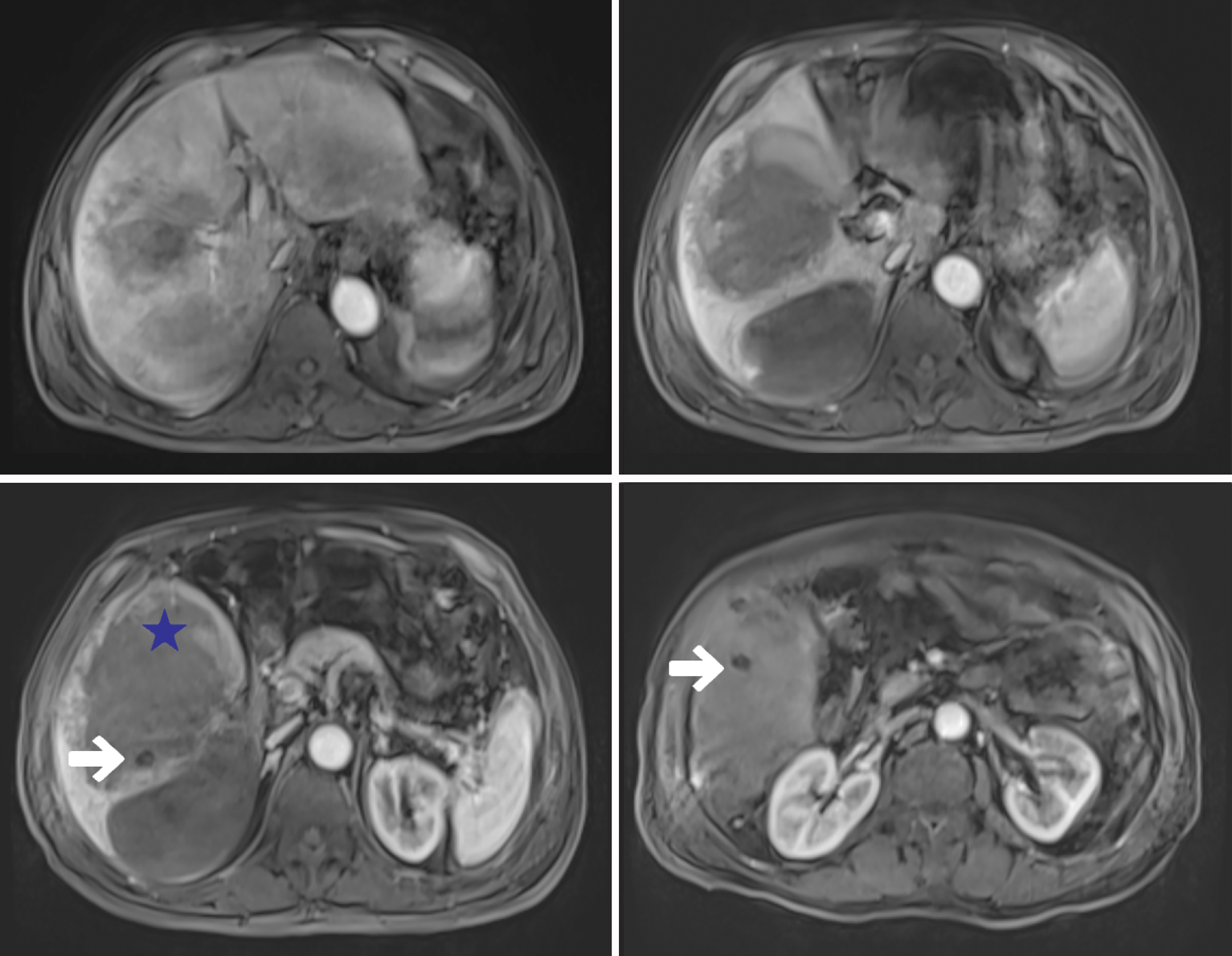
Figure 2 Magnetic resonance imaging of the abdomen showing a mass of mixed signal in the right lobe of the liver.
White arrow: Several rounded long T1 signals in the mixed-signal mass; Blue star: The “hole sign”.
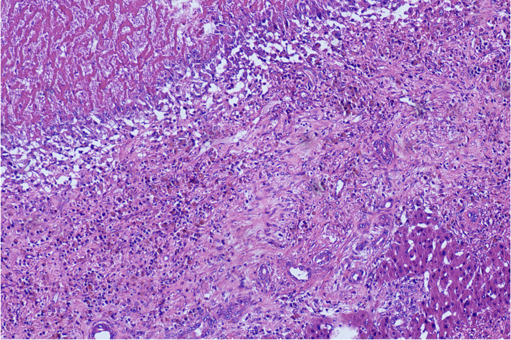
Figure 3 Ulceration of the gallbladder with fibrosis and inflammatory cell infiltration.
- Citation: Huang HW, Wang H, Leng C, Mei B. Formation and rupture of liver hematomas caused by intrahepatic gallbladder perforation: A case report and review of literature. World J Gastrointest Surg 2024; 16(10): 3301-3311
- URL: https://www.wjgnet.com/1948-9366/full/v16/i10/3301.htm
- DOI: https://dx.doi.org/10.4240/wjgs.v16.i10.3301


