Copyright
©The Author(s) 2023.
World J Gastrointest Surg. Sep 27, 2023; 15(9): 2089-2097
Published online Sep 27, 2023. doi: 10.4240/wjgs.v15.i9.2089
Published online Sep 27, 2023. doi: 10.4240/wjgs.v15.i9.2089
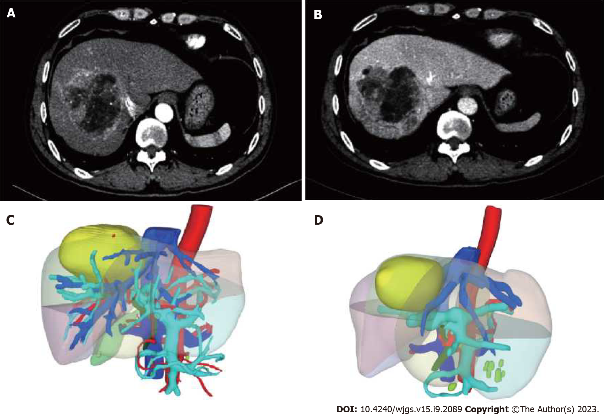
Figure 1 Image data of case 1.
A: Arterial phase; B: Portal stage; C and D: 3D imaging before and after treatment.
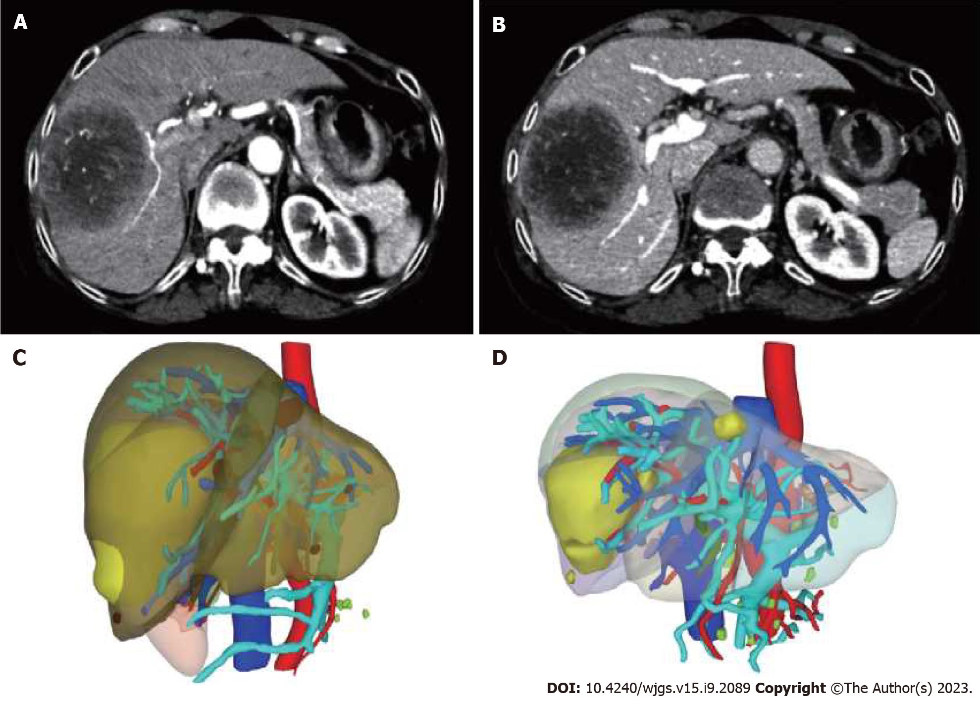
Figure 2 Image data of case 2.
A: Arterial phase; B: Portal stage; C and D: 3D imaging before and after treatment.
Figure 3 Complete laparoscopic right portal vein ligation surgery.
A: Dissection of the right branch of the portal vein within the Glisson's sheath at the first porta hepatis; B: Occlusion with titanium clip or ligation with No. 7 silk suture; C: Marking of the ischemic line on the surface of the liver with an electrocautery hook.
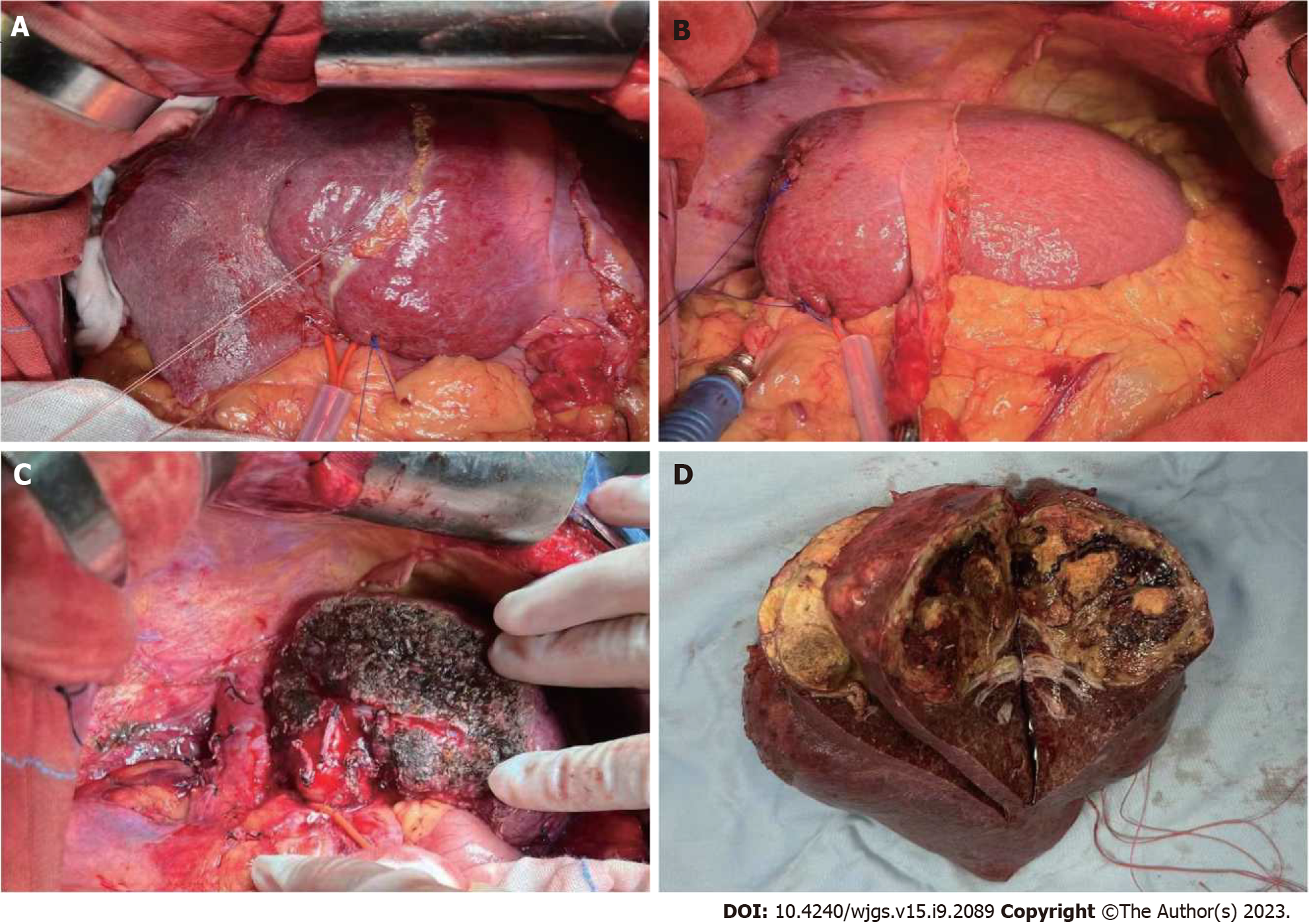
Figure 4 Right hemihepatectomy.
A: Intraoperative exploration revealed hypertrophied left lobe of the liver and a marked ischemic line; B: Postoperative left lobe of the liver; C: Surgical section; D: Tumor specimen.
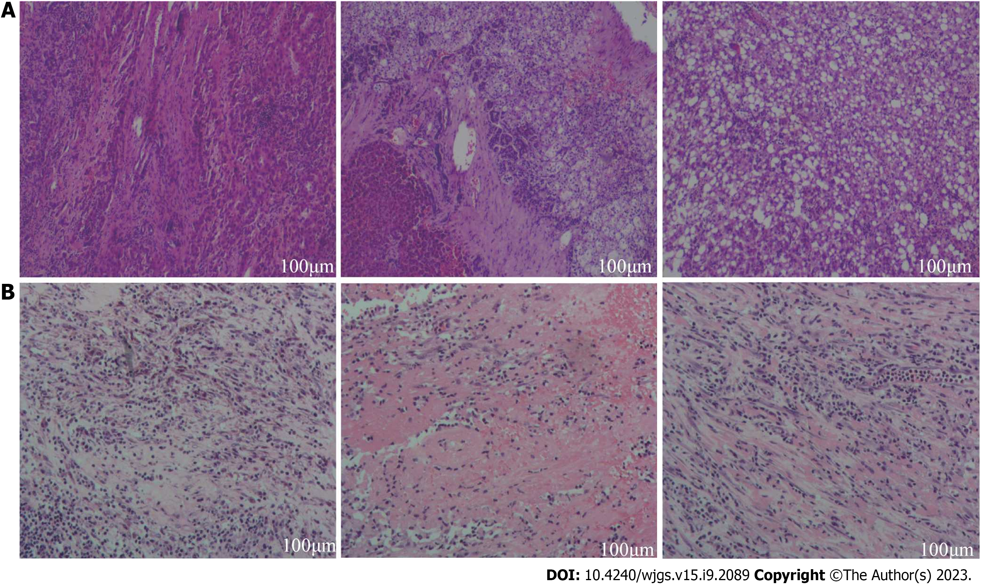
Figure 5 Histopathological image.
A: Case 1; B: Case 2.
- Citation: Gao Q, Zhu GZ, Han CY, Ye XP, Huang HS, Mo ST, Peng T. Dual transformation therapy for giant hepatocellular carcinoma: Two case reports and review of literature. World J Gastrointest Surg 2023; 15(9): 2089-2097
- URL: https://www.wjgnet.com/1948-9366/full/v15/i9/2089.htm
- DOI: https://dx.doi.org/10.4240/wjgs.v15.i9.2089


