Copyright
©The Author(s) 2023.
World J Gastrointest Surg. Jul 27, 2023; 15(7): 1532-1541
Published online Jul 27, 2023. doi: 10.4240/wjgs.v15.i7.1532
Published online Jul 27, 2023. doi: 10.4240/wjgs.v15.i7.1532
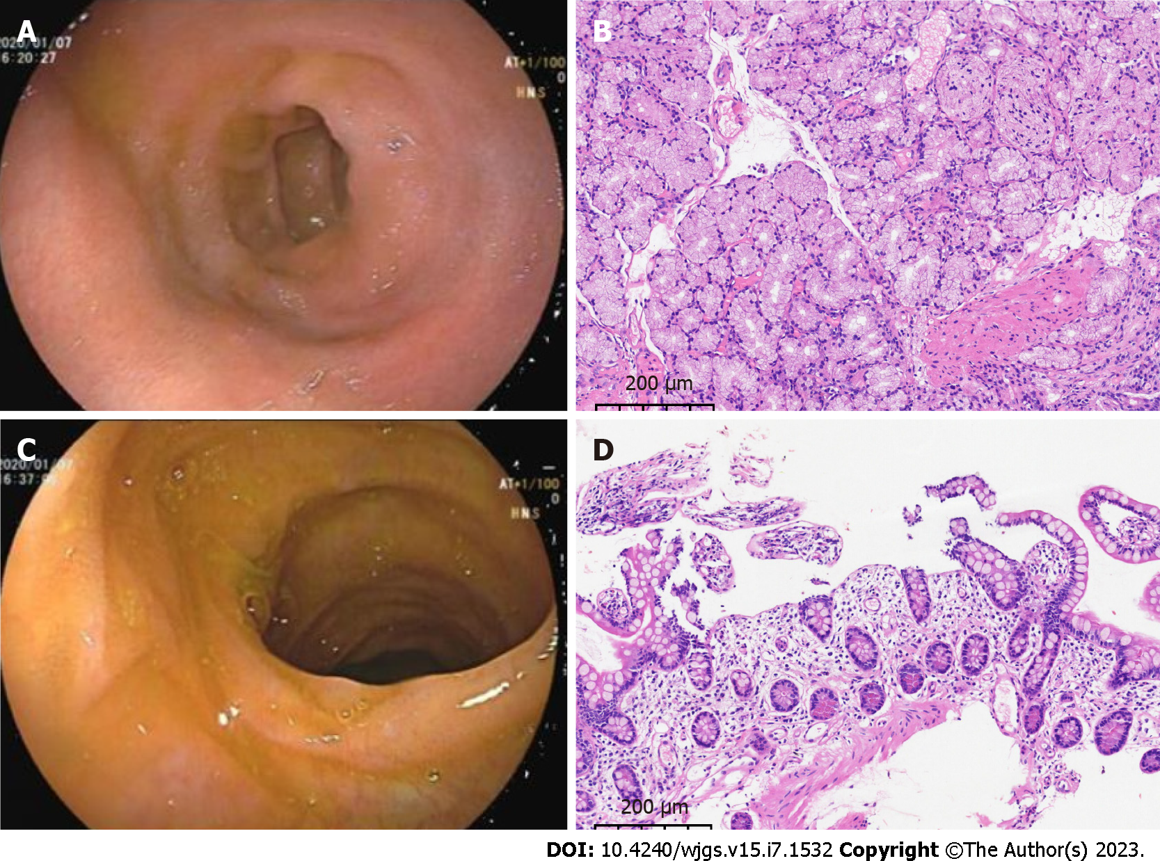
Figure 1 Gastrointestinal endoscopy and pathology.
A: Gastroduodenoscopy revealed an ulcerated scar on the duodenal bulb; B: Pathology of the intestinal mucosa of the duodenal bulb showed chronic inflammation with localized erosion (magnification, 10 ×); C: Painless colonoscopy showed no significant abnormalities in the mucosa of the terminal ileum; D: Pathology of the mucosa of the terminal ileum showed chronic inflammation (magnification 10 ×).
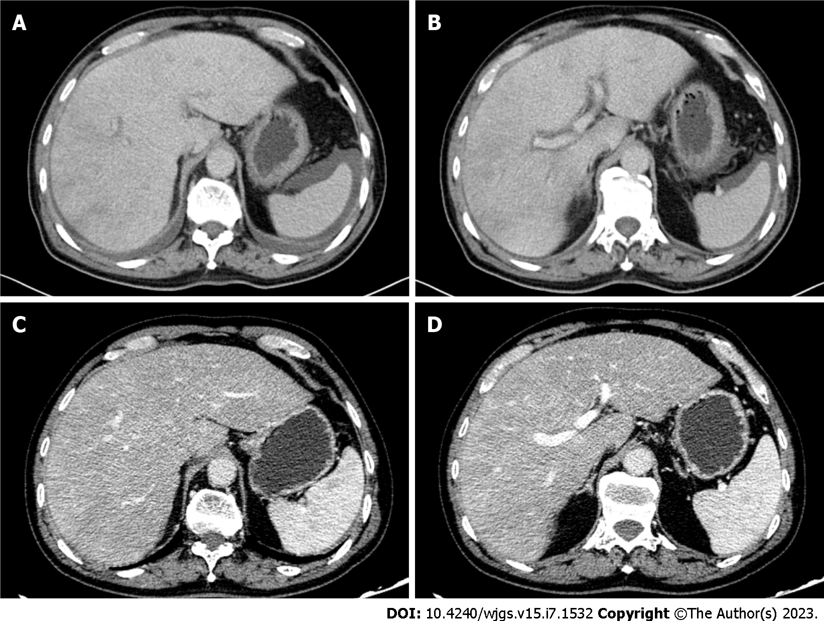
Figure 2 Contrast-enhanced computed tomography images of the upper abdomen of the patient.
A and B: Pretreatment enhanced computed tomography (CT) showed edema around the portal branch and fine compressed flattening of the inferior hepatic segment and hepatic veins with ascites; C and D: Post-treatment enhanced CT showed that the inferior vena cava and hepatic veins of the hepatic segment were thin, and congestion of the hepatic venules was improved.
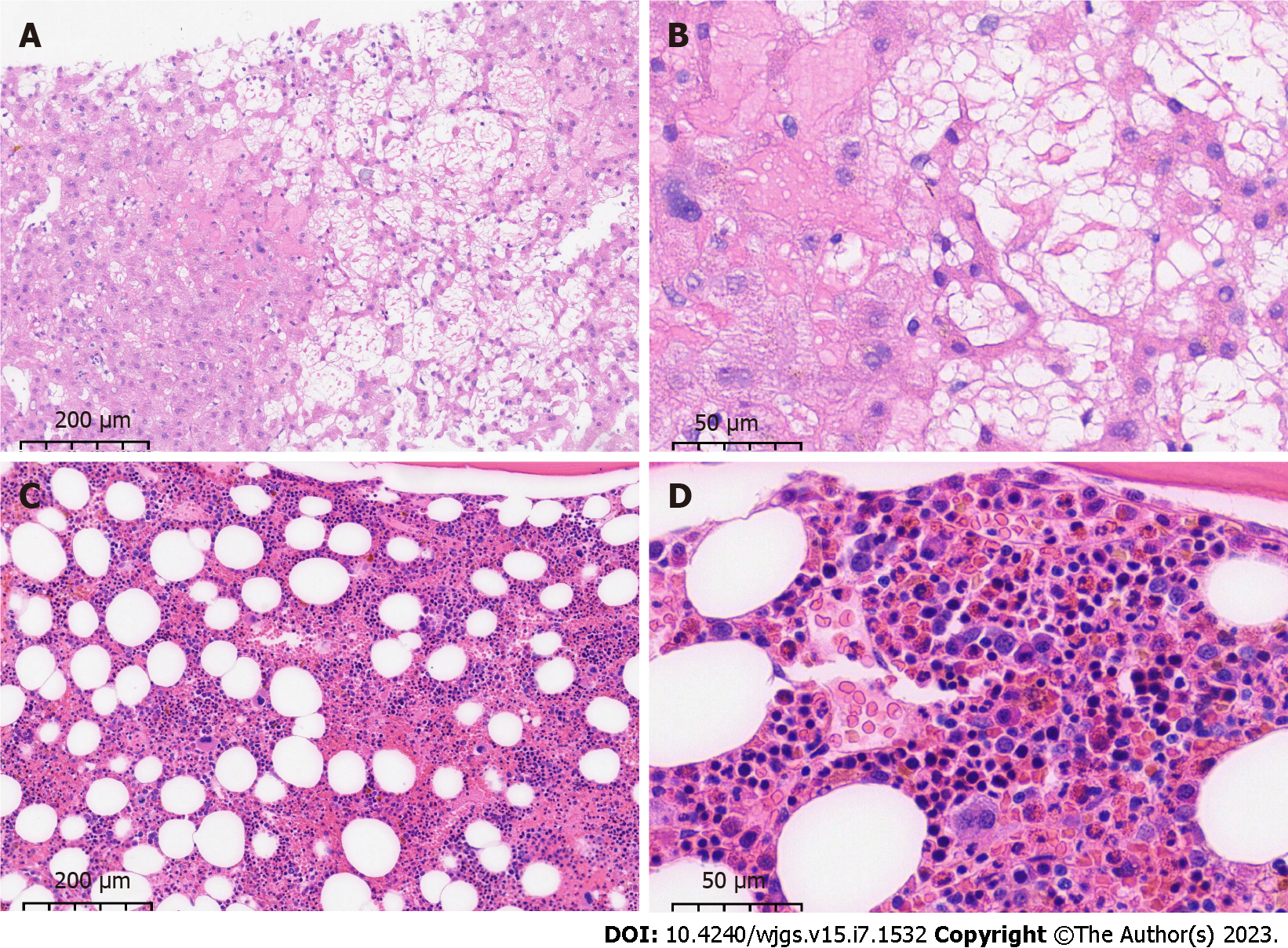
Figure 3 Pathology of liver and bone puncture of the patient.
A and B: Ultrasound-guided liver puncture biopsy suggested hepatic sinusoidal obstruction syndrome. Hepatic lobular structures, hydropic degeneration of hepatocytes, dilated and stagnant lamellar hepatic sinusoids with loss of hepatocytes, residual reticulofibrous scaffolds and insignificant inflammatory cell infiltration were seen (A: magnification 10 ×; B: magnification 40 ×); C and D: Bone marrow aspiration biopsy showed an active proliferation of bone marrow tissue but a significantly higher percentage of eosinophils (23%) with approximately normal morphology (C: magnification 10 ×; D: magnification 40 ×).
- Citation: Xu XT, Wang BH, Wang Q, Guo YJ, Zhang YN, Chen XL, Fang YF, Wang K, Guo WH, Wen ZZ. Idiopathic hypereosinophilic syndrome with hepatic sinusoidal obstruction syndrome: A case report and literature review. World J Gastrointest Surg 2023; 15(7): 1532-1541
- URL: https://www.wjgnet.com/1948-9366/full/v15/i7/1532.htm
- DOI: https://dx.doi.org/10.4240/wjgs.v15.i7.1532


