Copyright
©The Author(s) 2023.
World J Gastrointest Surg. Apr 27, 2023; 15(4): 553-565
Published online Apr 27, 2023. doi: 10.4240/wjgs.v15.i4.553
Published online Apr 27, 2023. doi: 10.4240/wjgs.v15.i4.553
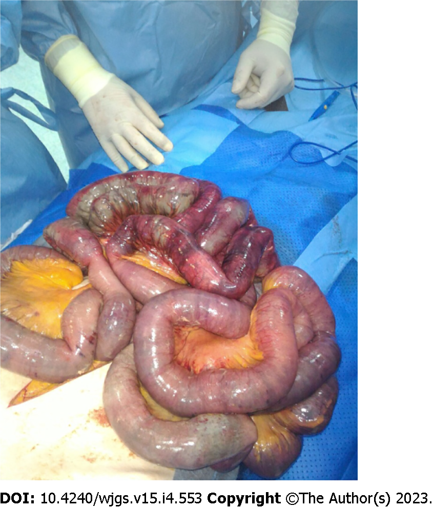
Figure 1 Intraoperative finding of diffuse ileal ischemia.
(Personal observation).
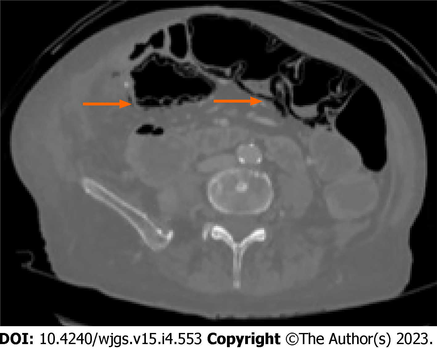
Figure 2 Computed tomography-scan with evidence of cystoid or bubble-like pattern pneumatosis intestinalis, identified by the orange arrow.
(Personal observation).
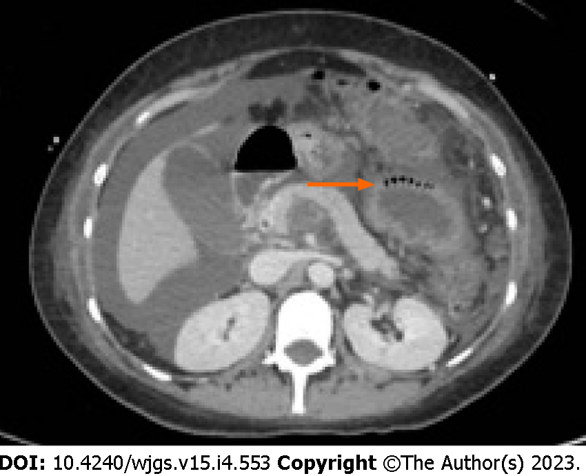
Figure 3 Computed tomography-scan documenting a linear pattern at the level of the colonic wall, identified by the orange arrow.
(Personal observation).
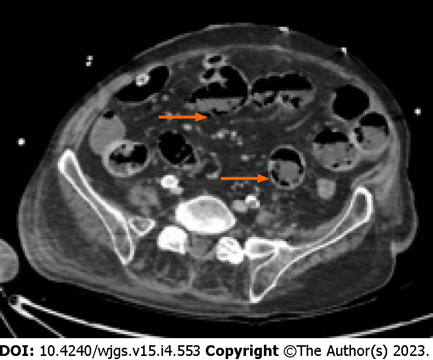
Figure 4 Computed tomography-scan documenting circumferential pattern pneumatosis intestinalis, identified by the orange arrow.
(Personal observation).
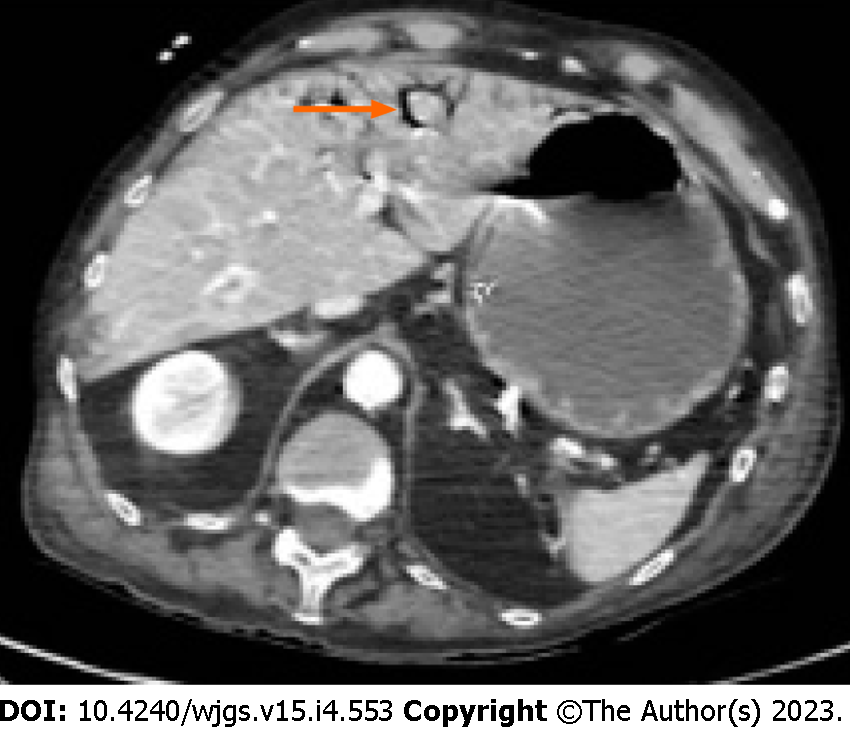
Figure 5 Computed tomography-scan documenting localized portal venous gas, identified by the orange arrow.
(Personal observation).
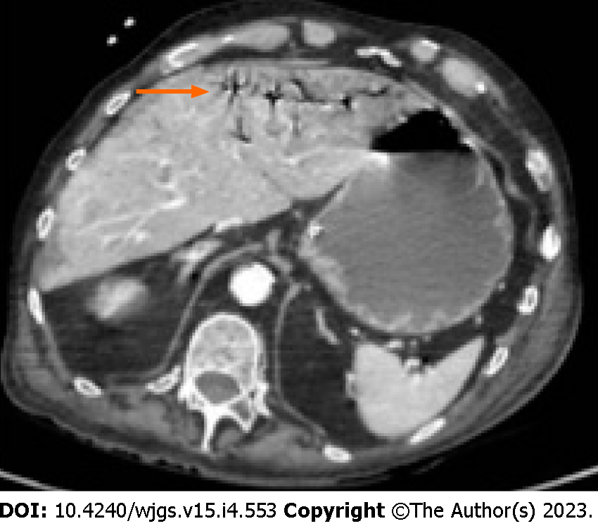
Figure 6 Computed tomography-scan documenting diffuse portal venous gas, identified by the orange arrow.
(Personal observation).
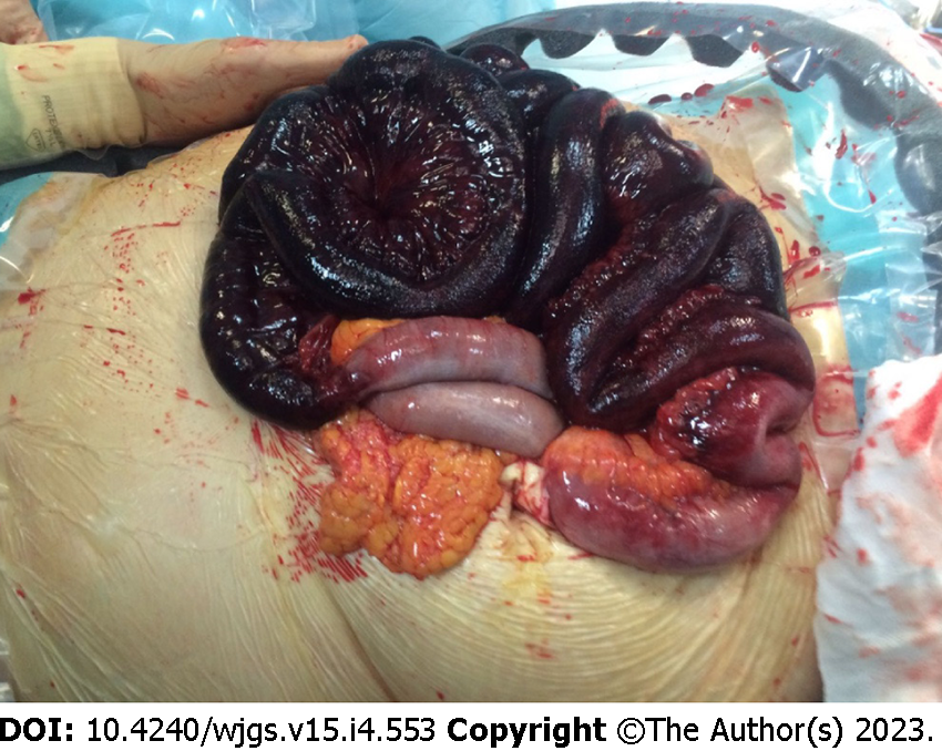
Figure 7 Intraoperative finding of transmural infarction with intestinal necrosis.
(Personal observation).
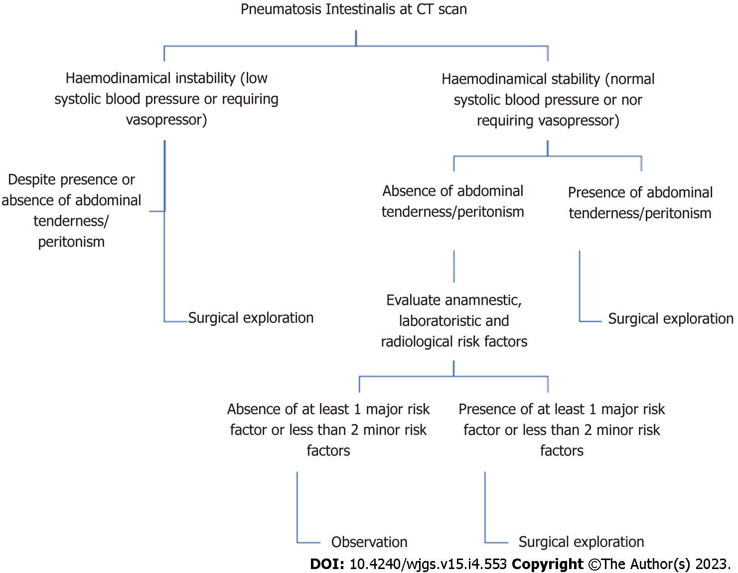
Figure 8 Algorithm to guide clinical decisions in patients with pneumatosis intestinalis.
CT: Computed tomography.
- Citation: Tropeano G, Di Grezia M, Puccioni C, Bianchi V, Pepe G, Fico V, Altieri G, Brisinda G. The spectrum of pneumatosis intestinalis in the adult. A surgical dilemma. World J Gastrointest Surg 2023; 15(4): 553-565
- URL: https://www.wjgnet.com/1948-9366/full/v15/i4/553.htm
- DOI: https://dx.doi.org/10.4240/wjgs.v15.i4.553


