Copyright
©The Author(s) 2023.
World J Gastrointest Surg. Oct 27, 2023; 15(10): 2376-2381
Published online Oct 27, 2023. doi: 10.4240/wjgs.v15.i10.2376
Published online Oct 27, 2023. doi: 10.4240/wjgs.v15.i10.2376
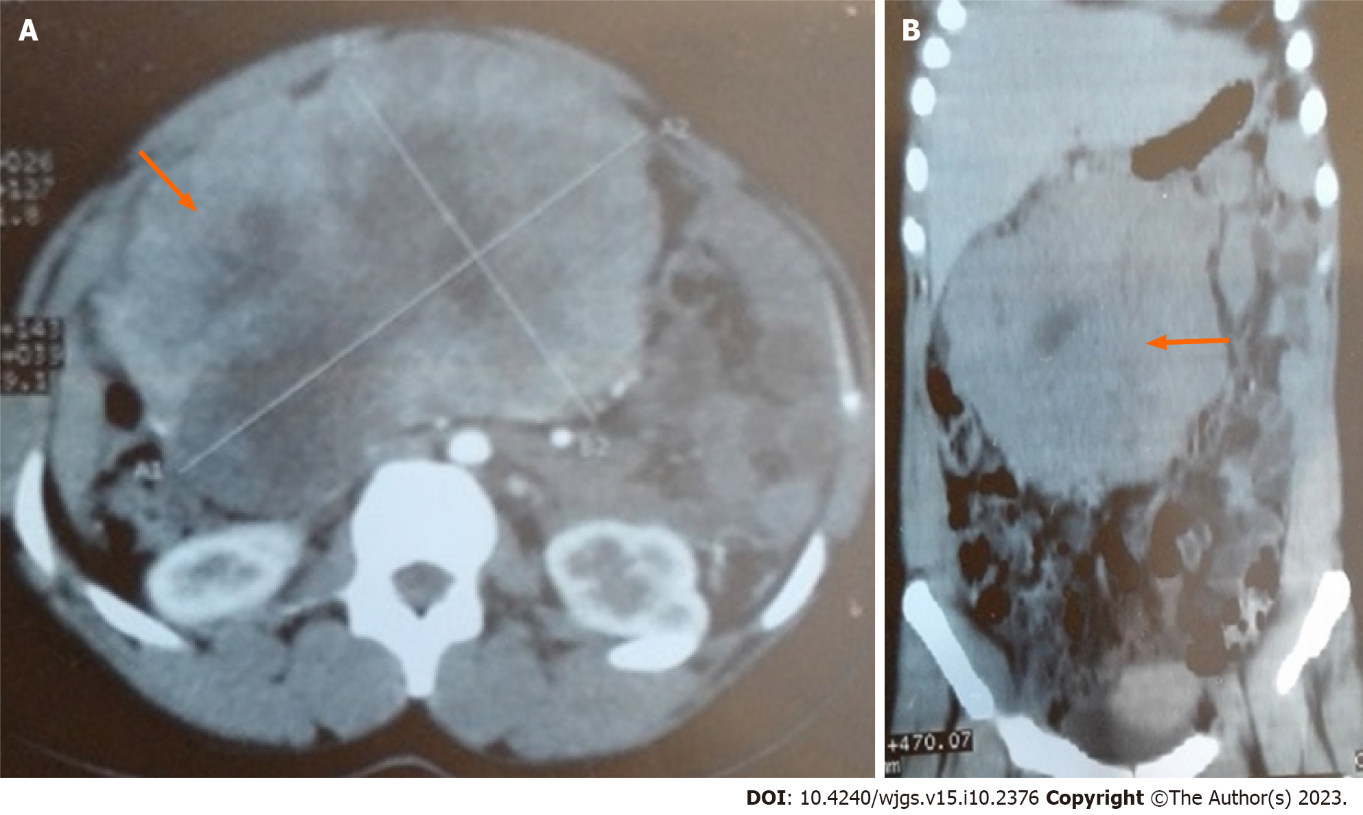
Figure 1 Abdominal computed tomography showed a huge heterogeneous mesentery mass.
A: Transverse view; B: Coronal view.
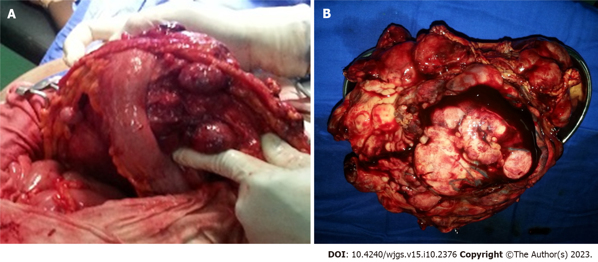
Figure 2 Operatives images of this case report.
A: Intraoperative procedure found multi-lobulated mass of gastrocolic ligament; B: Macroscopic aspect of removed giant mass of gastrocolic ligament.
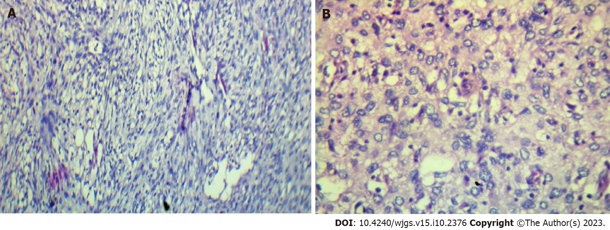
Figure 3 Histopathological analysis.
Immunohistochemical results show that the tumours cells are positive for Bap1 (A), p16 (A) and MDM2 (B) and negative for actin, desmin, calretinin, PS100, melana A, ERG, p63, cytokeratin AE1/AE3, EMA, chromogranin A and CDK4. A: A population of fusiform cells (× 200); B: A population of epithelioid cells and atypical adipose cells in a myxoid and vascularized matrix (× 200).
- Citation: Kassi ABF, Yenon KS, Kassi FMH, Adjeme AJ, Diarra KM, Bombet-Kouame C, Kouassi M. Giant dedifferentiated liposarcoma of the gastrocolic ligament: A case report. World J Gastrointest Surg 2023; 15(10): 2376-2381
- URL: https://www.wjgnet.com/1948-9366/full/v15/i10/2376.htm
- DOI: https://dx.doi.org/10.4240/wjgs.v15.i10.2376


