Copyright
©The Author(s) 2023.
World J Gastrointest Surg. Oct 27, 2023; 15(10): 2357-2361
Published online Oct 27, 2023. doi: 10.4240/wjgs.v15.i10.2357
Published online Oct 27, 2023. doi: 10.4240/wjgs.v15.i10.2357
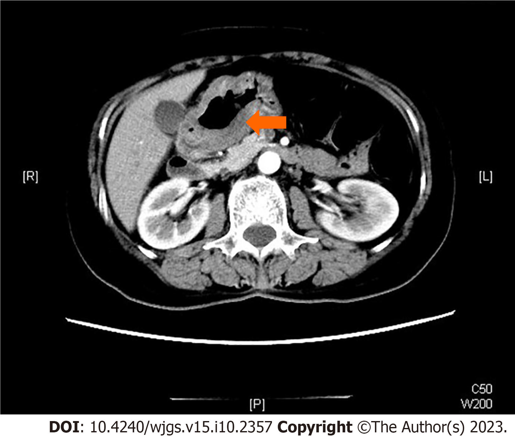
Figure 1 Enhanced computed tomography examination of the abdomen.
Invasive lesion close to the pylorus can be seen, and the edges between the tumor and peripheral organs such as the pancreas and liver are still clear (arrow).
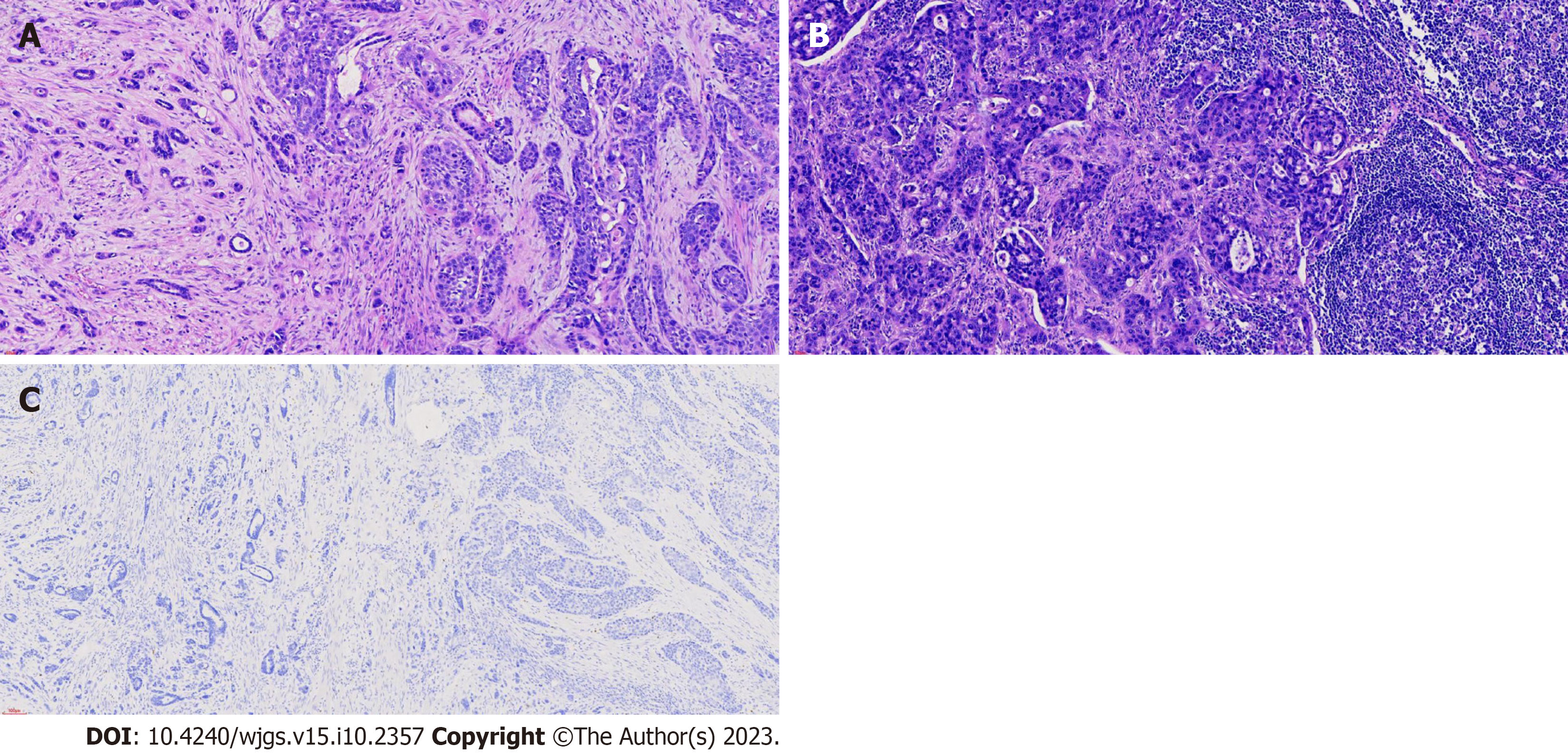
Figure 2 Histopathological analysis and immunohistochemical examination.
A and B: Histopathological analysis and immunohistochemical examination of the resected specimen. Gastric adenosquamous carcinoma (20 ×), lymph node metastasis (20 ×); C: Immunohistochemistry staining for alpha-fetoprotein (AFP) of the resected specimen. Immunohistochemistry staining for AFP (10 ×).
- Citation: Sun L, Wei JJ, An R, Cai HY, Lv Y, Li T, Shen XF, Du JF, Chen G. Gastric adenosquamous carcinoma with an elevated serum level of alpha-fetoprotein: A case report. World J Gastrointest Surg 2023; 15(10): 2357-2361
- URL: https://www.wjgnet.com/1948-9366/full/v15/i10/2357.htm
- DOI: https://dx.doi.org/10.4240/wjgs.v15.i10.2357


