Copyright
©The Author(s) 2022.
World J Gastrointest Surg. Sep 27, 2022; 14(9): 918-929
Published online Sep 27, 2022. doi: 10.4240/wjgs.v14.i9.918
Published online Sep 27, 2022. doi: 10.4240/wjgs.v14.i9.918
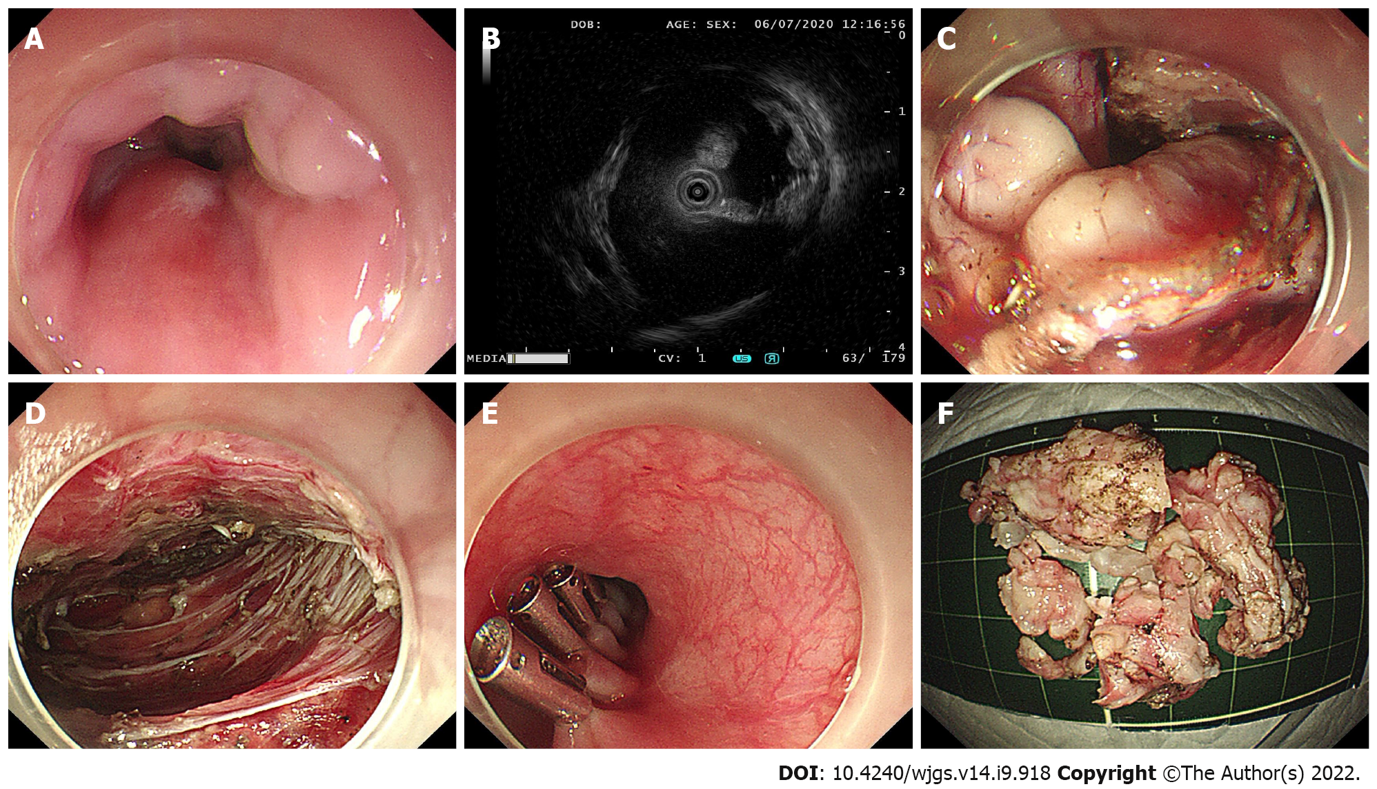
Figure 1 The procedure of submucosal tunneling endoscopic resection.
A: Endoscopic view of the tumor; B: Endoscopic ultrasonography view of the tumor; C: The submucosal tumor exposed using the submucosal tunnel technique; D: Endoscopic view of the submucosal tunnel after the tumor was removed; E: The mucosal entry closed by clips; F: The piecemeal resected tumor.
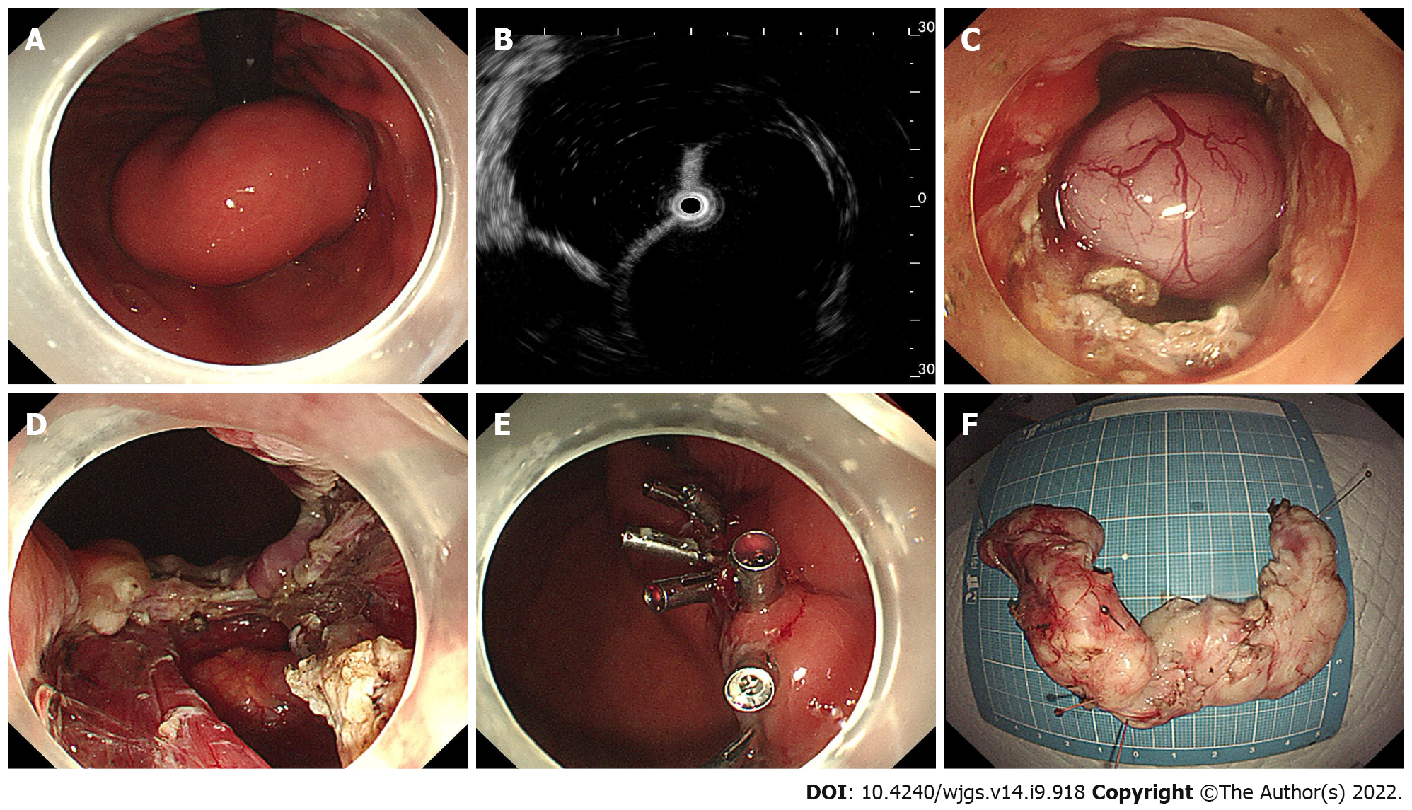
Figure 2 Case illustration of endoscopic full-thickness resection.
A: Endoscopic view of the tumor; B: Endoscopic ultrasonography view of the tumor; C: The submucosal tumor exposed by full-thickness resection; D: The wound surface after removal of the tumor; E: The gastric wall defect was closed with endo-clips; F: The horseshoe-shaped specimen.
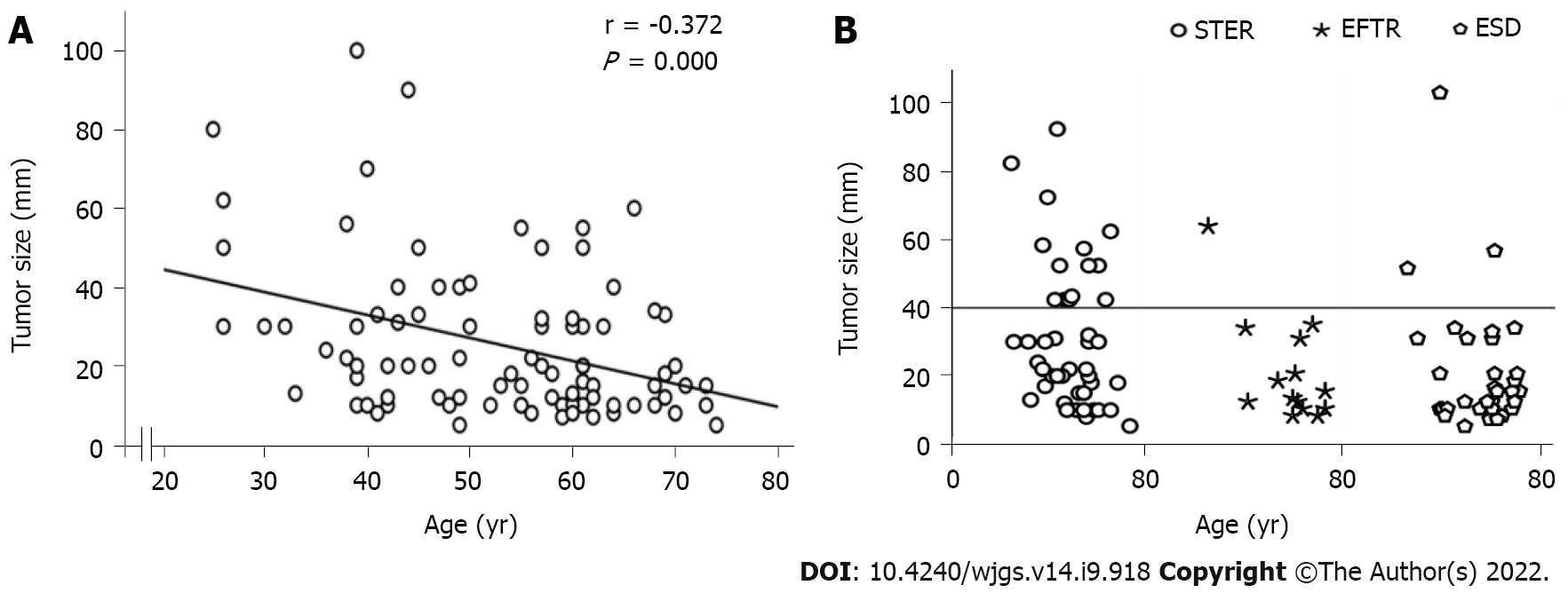
Figure 3 Tumor size.
A: There was a significant negative correlation between age and tumor size; B: Tumor size at different ages in the submucosal tunneling endoscopic resection group, endoscopic full-thickness resection group and endoscopic submucosal dissection group are shown. The circle dots above the horizontal line represent tumors larger than 4 cm. STER: Submucosal tunneling endoscopic resection; EFTR: Endoscopic full-thickness resection; ESD: Endoscopic submucosal dissection.
- Citation: Wang YP, Xu H, Shen JX, Liu WM, Chu Y, Duan BS, Lian JJ, Zhang HB, Zhang L, Xu MD, Cao J. Predictors of difficult endoscopic resection of submucosal tumors originating from the muscularis propria layer at the esophagogastric junction. World J Gastrointest Surg 2022; 14(9): 918-929
- URL: https://www.wjgnet.com/1948-9366/full/v14/i9/918.htm
- DOI: https://dx.doi.org/10.4240/wjgs.v14.i9.918


