Copyright
©The Author(s) 2022.
World J Gastrointest Surg. Sep 27, 2022; 14(9): 1082-1085
Published online Sep 27, 2022. doi: 10.4240/wjgs.v14.i9.1082
Published online Sep 27, 2022. doi: 10.4240/wjgs.v14.i9.1082
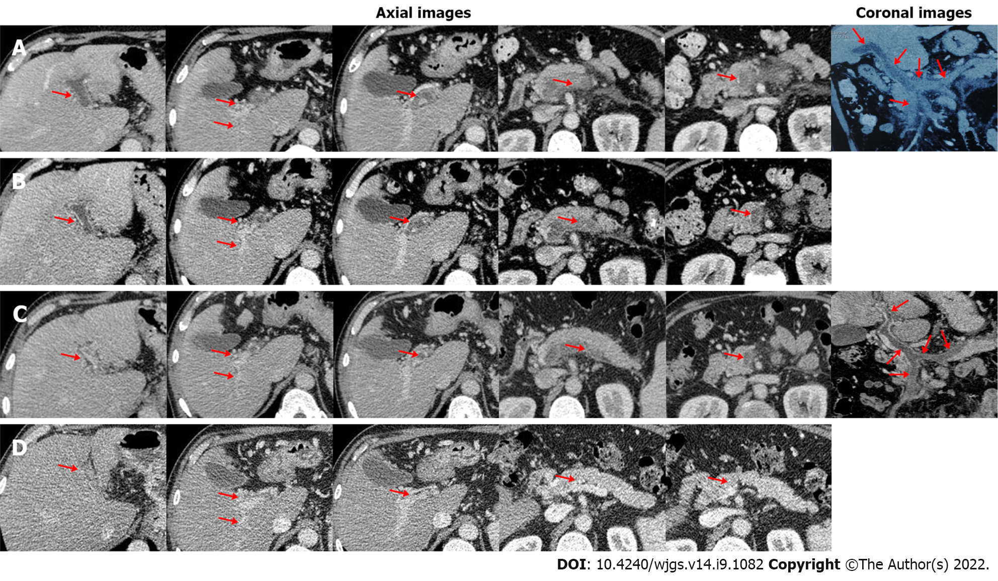
Figure 1 Axial and coronal computed tomography images in this patient.
A: On day 1 of admission, computed tomography (CT) images demonstrated occlusive thrombosis within the main portal vein (MPV), left portal vein (LPV), right portal vein (RPV), confluence of the superior mesenteric vein (SMV) and splenic vein (SV), SMV, and SV, with fine collaterals around the hilum (red arrow); B: On day 5, CT images demonstrated partially recanalized LPV and RPV (red arrow); C: On day 10, CT images demonstrated partially recanalized MPV, LPV, and RPV (red arrow); D: After 5-mo anticoagulation with rivaroxaban, CT images demonstrated completely recanalized SMV and SV (red arrow).
- Citation: Gao FB, Wang L, Zhang WX, Shao XD, Guo XZ, Qi XS. Successful treatment of acute symptomatic extensive portal venous system thrombosis by 7-day systemic thrombolysis. World J Gastrointest Surg 2022; 14(9): 1082-1085
- URL: https://www.wjgnet.com/1948-9366/full/v14/i9/1082.htm
- DOI: https://dx.doi.org/10.4240/wjgs.v14.i9.1082


