Copyright
©The Author(s) 2022.
World J Gastrointest Surg. Nov 27, 2022; 14(11): 1285-1296
Published online Nov 27, 2022. doi: 10.4240/wjgs.v14.i11.1285
Published online Nov 27, 2022. doi: 10.4240/wjgs.v14.i11.1285
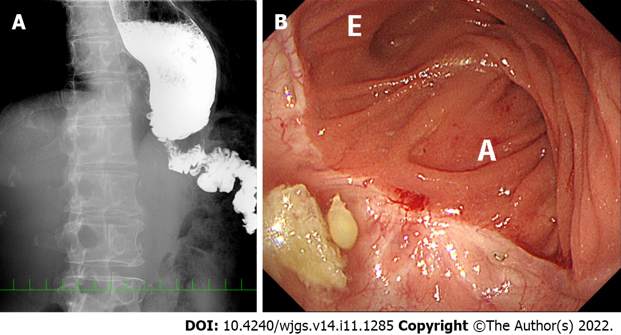
Figure 1 Stricture of the jejunal limb before reoperation in case 5.
A: Preoperative X-ray examination with contrast medium shows poor passage of contrast medium at the hiatus, jejunal stenosis (approximately 1 cm in length) and jejunal bending at the hiatus, in addition to distal esophageal dilatation; B: Endoscopic luminal examination shows an intact anastomosis and a bent or tortuous efferent jejunal limb, but the scope could be passed through this portion. E: Efferent side; A: Afferent side.
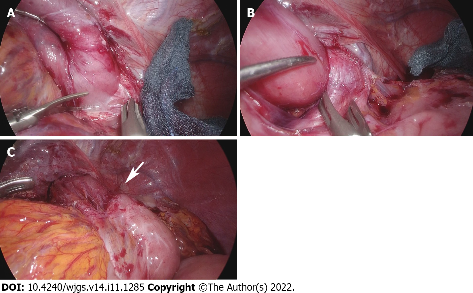
Figure 2 Stricture of the jejunal limb during reoperation in case 5.
A: Fibrous adhesions are observed around the hiatus; B: Loose adhesions are present on the left crus; C: A pressure mark (white arrow) is identified on the jejunal limb after completion of adhesiolysis up to the anastomosis.
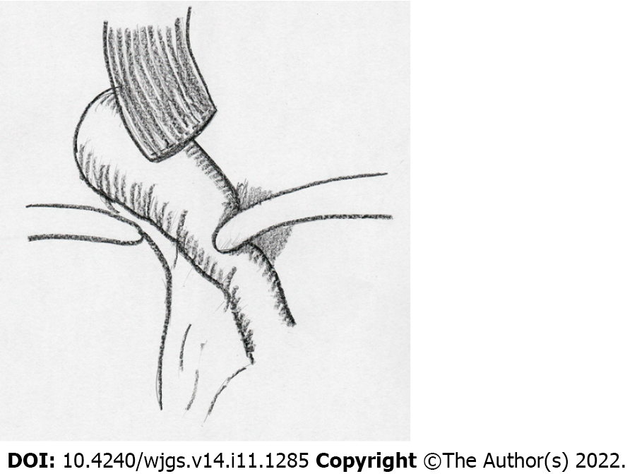
Figure 3 Schema of the complicated disorder after overlapped esophagojejunostomy following laparoscopic total gastrectomy.
The jejunal limb stricture was resulted from the shortened remnant esophagus and jejunal bending resulting from loose and fibrous adhesions on the left crus at the esophageal hiatus.
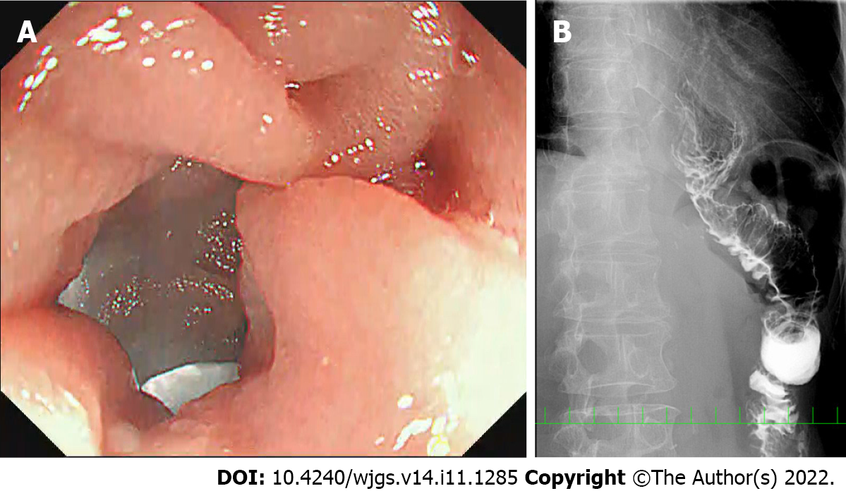
Figure 4 No stricture of the jejunal limb on intra- or postoperative examinations in case 5.
A: Intraoperative endoscopic luminal examination showed a straight efferent jejunal limb; B: Postoperative X-ray examination with contrast medium showed good passage at the hiatus, as well as improvement of jejunal limb bending and distal esophageal dilatation.
- Citation: Noshiro H, Okuyama K, Yoda Y. Disturbed passage of jejunal limb near esophageal hiatus after overlapped esophagojejunostomy following laparoscopic total gastrectomy. World J Gastrointest Surg 2022; 14(11): 1285-1296
- URL: https://www.wjgnet.com/1948-9366/full/v14/i11/1285.htm
- DOI: https://dx.doi.org/10.4240/wjgs.v14.i11.1285


