Copyright
©The Author(s) 2021.
World J Gastrointest Surg. Jan 27, 2021; 13(1): 76-86
Published online Jan 27, 2021. doi: 10.4240/wjgs.v13.i1.76
Published online Jan 27, 2021. doi: 10.4240/wjgs.v13.i1.76
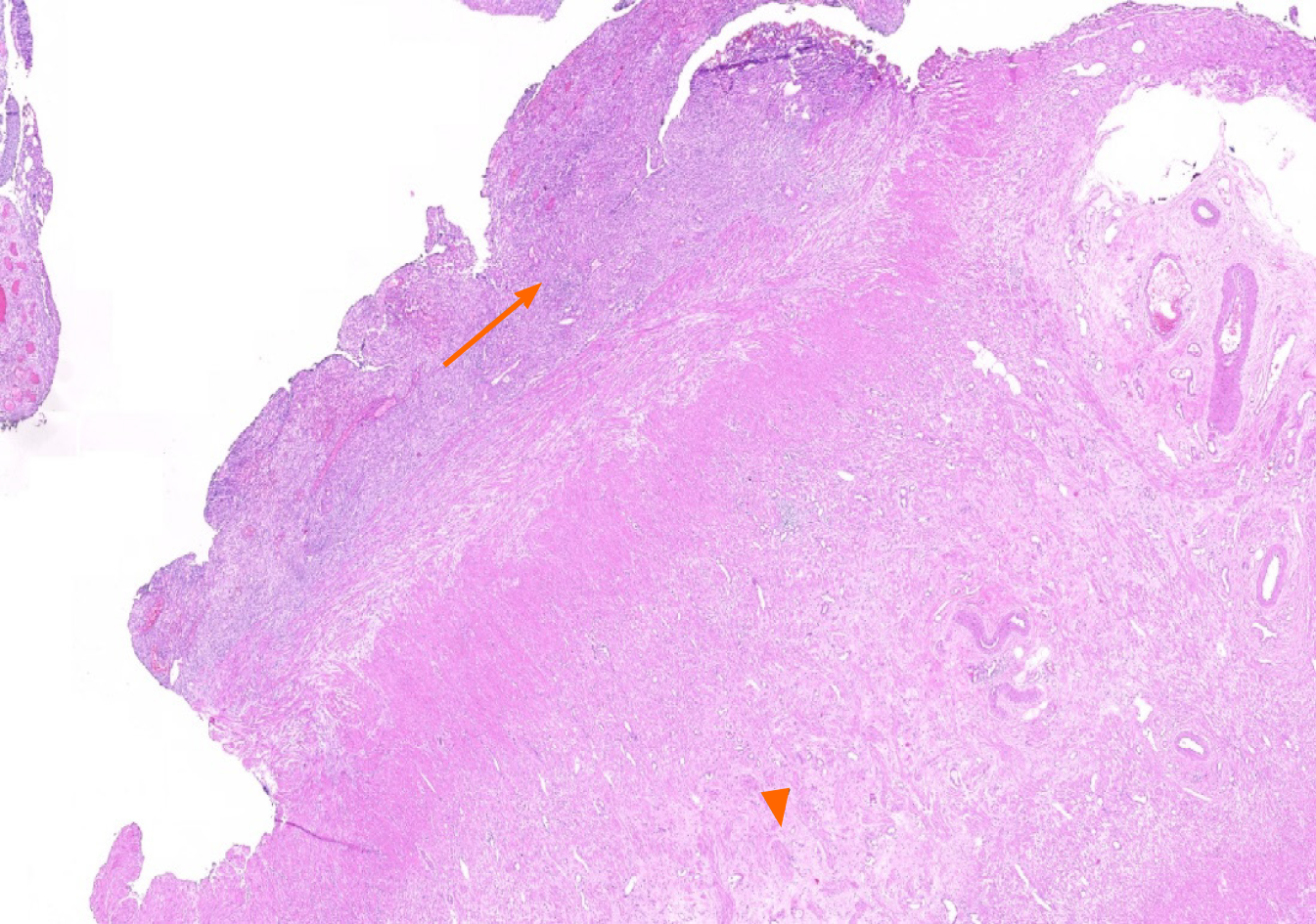
Figure 1 Fibrous obliteration of appendix vermiformis (arrow head), acute and chronic inflammatory cell infiltration (arrow) within the appendix wall and subserosal fatty tissue (HE × 10).
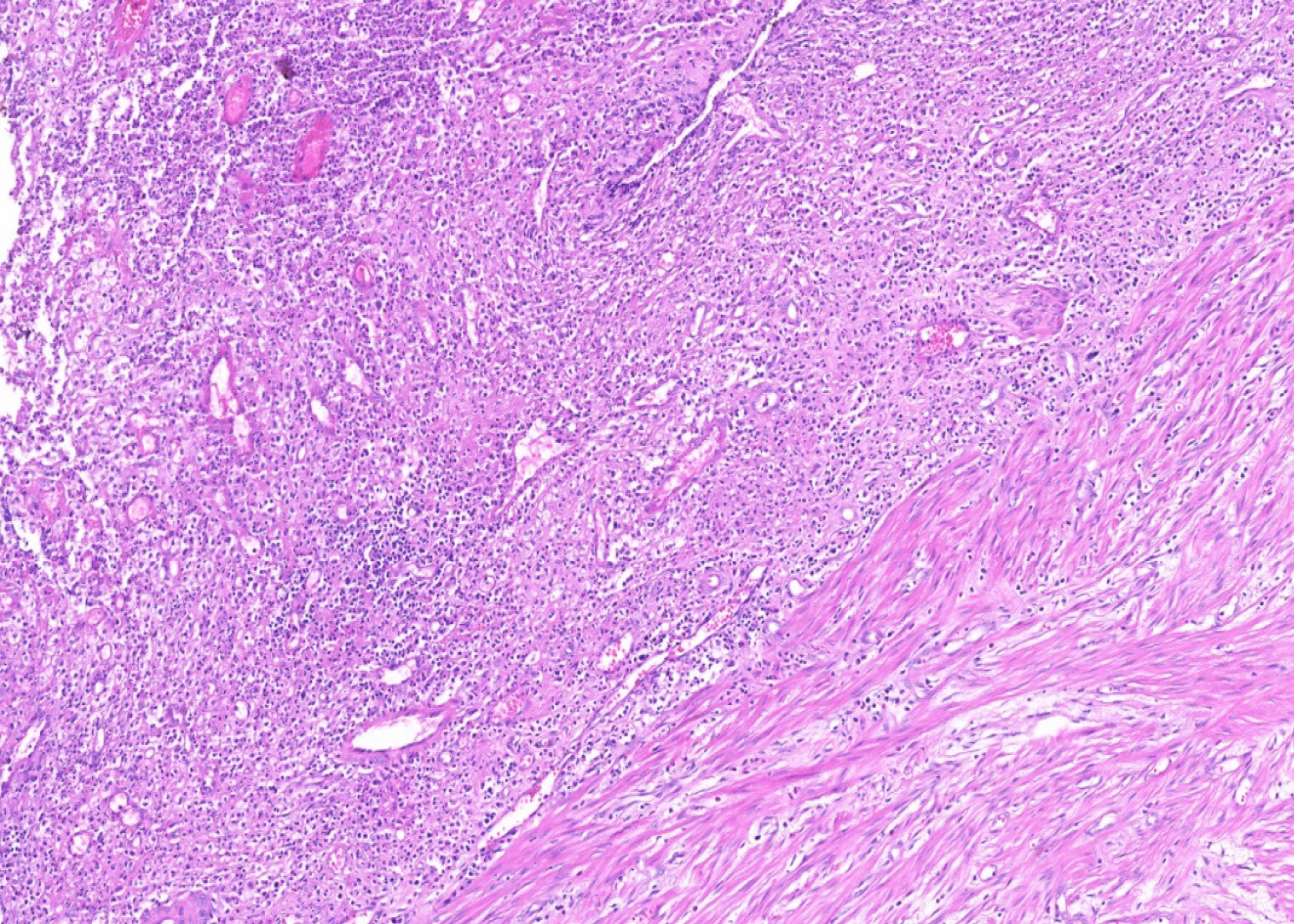
Figure 2 Xanthogranulomatous inflammation (a mixture of macrophages, lymphocytes, plasma cells, and neutrophils) (HE × 50).
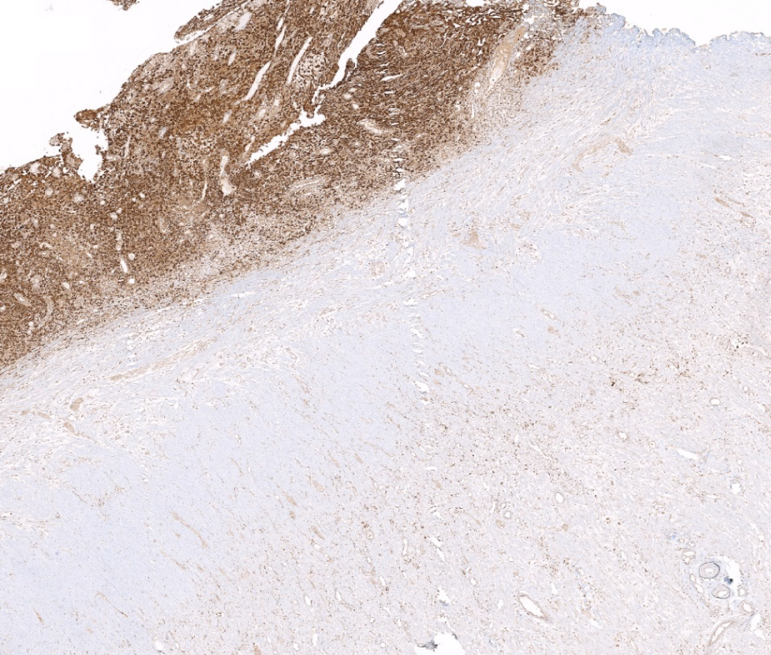
Figure 3 Macrophages showing positive staining for CD68 antibody.
- Citation: Akbulut S, Demyati K, Koc C, Tuncer A, Sahin E, Ozcan M, Samdanci E. Xanthogranulomatous appendicitis: A comprehensive literature review. World J Gastrointest Surg 2021; 13(1): 76-86
- URL: https://www.wjgnet.com/1948-9366/full/v13/i1/76.htm
- DOI: https://dx.doi.org/10.4240/wjgs.v13.i1.76


