Copyright
©The Author(s) 2019.
World J Gastrointest Surg. Feb 27, 2019; 11(2): 85-92
Published online Feb 27, 2019. doi: 10.4240/wjgs.v11.i2.85
Published online Feb 27, 2019. doi: 10.4240/wjgs.v11.i2.85
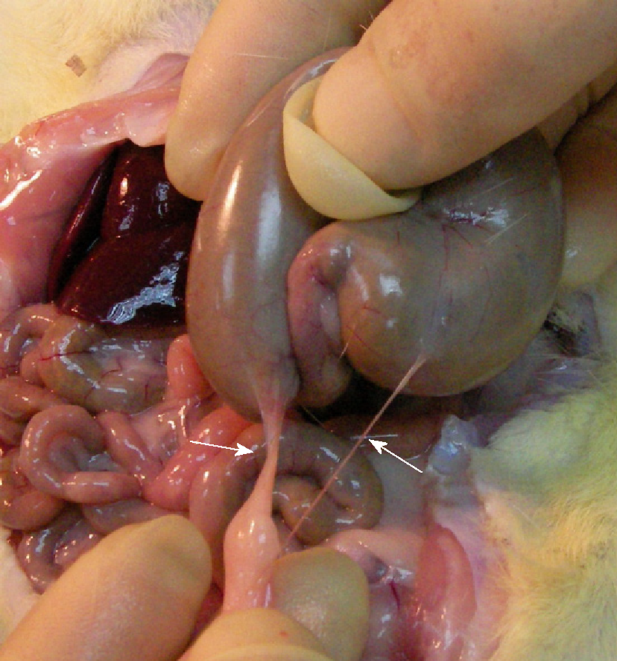
Figure 1 Representative photography of peritoneal adhesion in rats received abrasion injury on the surface of cecum.
Adhesion bands (arrows) were mostly found on apical region of the cecum with the adjacent omemtum.
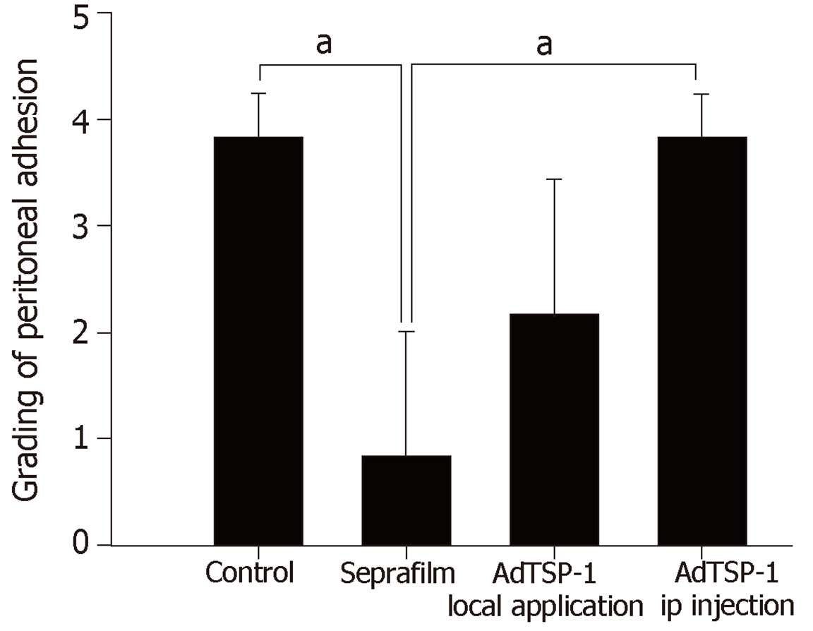
Figure 2 Quantifications of the peritoneal adhesion after experimental cecal abrasion injury.
Compared with controls (no treatment; group I), the application of Seprafilm (group II) significantly reduced the adhesion score and local administration of AdTSP-1 (group III) on the injured cecum the also attenuated the severity of peritoneal adhesion score. However, systemic delivery of AdTSP-1 (group IV) did not affect the formation of adhesion. Data were analyzed using the Kruskal-Wallis test, and are presented as mean ± SD. aP < 0.05, Seprafilm vs control; Seprafilm vs AdTSP-1, n = 10 in each group. AdTSP-1: Adenoviral vectors encoding mouse thrombospondin 1.
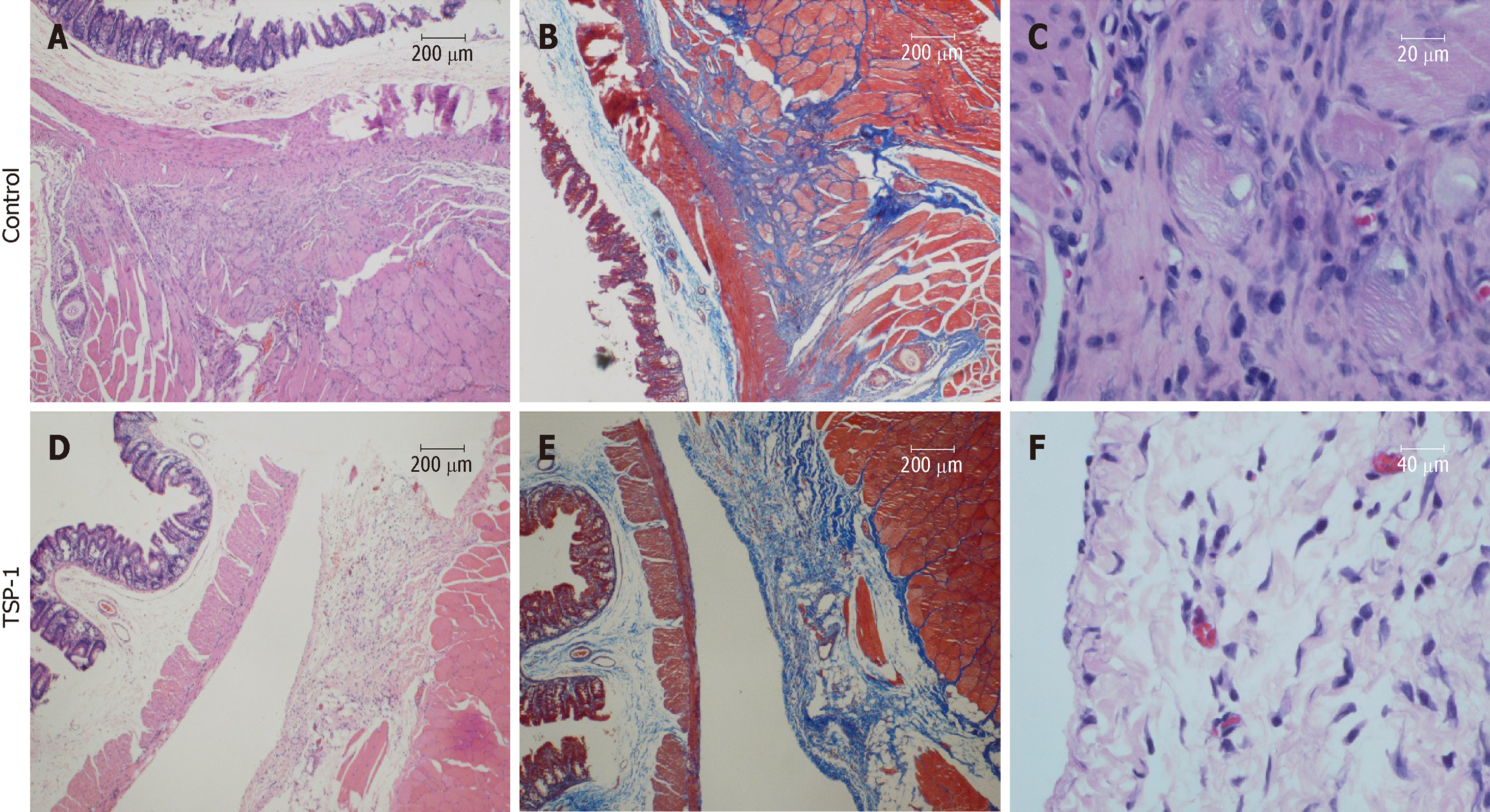
Figure 3 Representative histological sections of cecum with adhesion bands.
A: The muscularis propria is tightly adhered to the skeletal muscle by thick fibrotic tissue and infiltration of mononuclear cells (HE stain, 40 ×); B: The Masson trichrome stain indicates the intersecting of collagen-rich soft-tissue bands formed between the muscle layers (40 ×); C: HE stain shows the proliferative fibroblasts, increased collagen deposition and degenerative skeletal muscles (400 ×); D: Overexpressed TSP-1 on the injured cecum improved the formation of adhesion by separating the intestinal wall and skeletal muscle with a thin layer of loose connective tissue (HE, 40 ×); E: Masson trichrome stain identifies the loose connective tissue beside the skeletal muscle; F: Significantly fewer mononuclear cells infiltration in the loose connective tissue (HE, 400 ×). TSP-1: Thrombospondin 1.
- Citation: Tai YS, Jou IM, Jung YC, Wu CL, Shiau AL, Chen CY. In vivo expression of thrombospondin-1 suppresses the formation of peritoneal adhesion in rats. World J Gastrointest Surg 2019; 11(2): 85-92
- URL: https://www.wjgnet.com/1948-9366/full/v11/i2/85.htm
- DOI: https://dx.doi.org/10.4240/wjgs.v11.i2.85


