Copyright
©2011 Baishideng Publishing Group Co.
World J Diabetes. Apr 15, 2011; 2(4): 54-58
Published online Apr 15, 2011. doi: 10.4239/wjd.v2.i4.54
Published online Apr 15, 2011. doi: 10.4239/wjd.v2.i4.54
Figure 1 Staining by hematoxylin-eosin in embryonic pancreas exhibited pancreatic primary duct at 6 wk gestation.
A: The first-order bifurcation; B: The second-order bifurcation; C: The third-order bifurcation. Arrows show the duct beginning bifurcation. Bars = 200 μm.
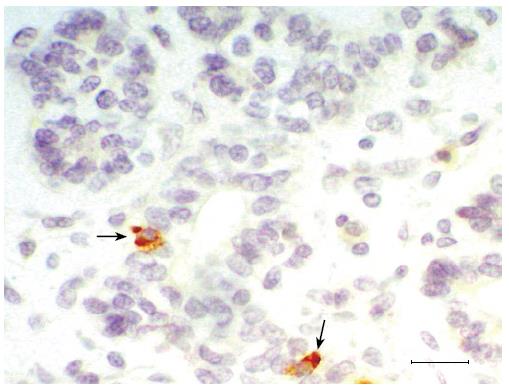
Figure 2 Expression of insulin in fetal pancreas.
Immunohistochemical staining is shown for insulin in pancreatic duct epithelium of 11 wk gestation. Arrow shows β-cell has differentiated before migration from duct. Bars = 50 μm.
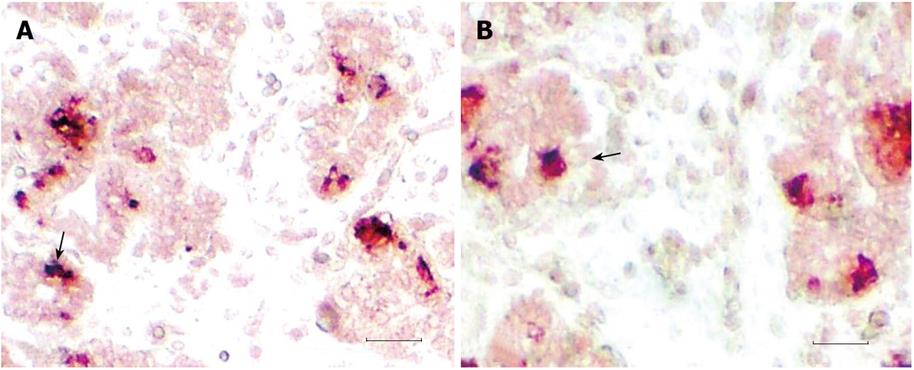
Figure 3 Immunohistochemical double staining in fetal pancreas.
A: For insulin glucagon (indigo) and insulin (scarlet); B: For somatostatin (indigo) Insulin (scarlet). Arrows showing the duct epithelia cell co-expressing insulin, glucagon or somatostatin at 11 wk gestation. Bars = 200 μm.
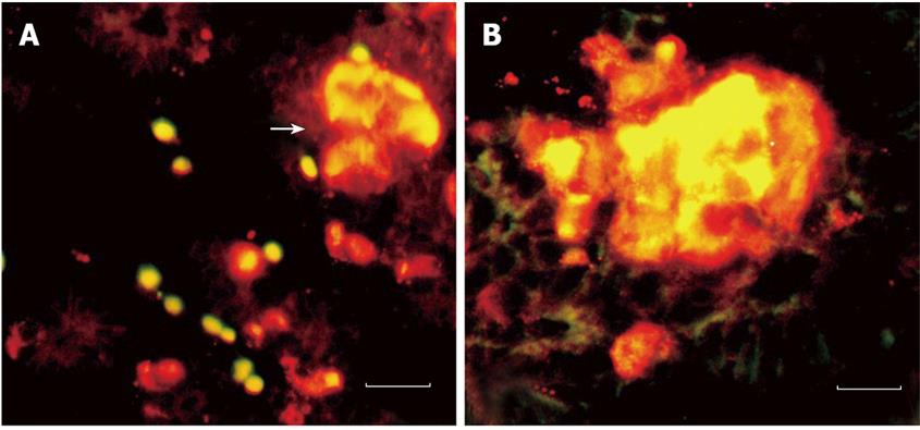
Figure 4 Double immunofluorescence staining in fetal pancreas insulin (FITC label, green) and nestin (Cy3 label, red).
A: At 11 wk, bar = 100 μm; B: At 14 wk, bar = 100 μm. Arrow shows that islet has formed and insulin-producing cells express nestin.
- Citation: Yang KM, Yong W, Li AD, Yang HJ. Insulin-producing cells are bi-potential and differentiatorsprior to proliferation in early human development. World J Diabetes 2011; 2(4): 54-58
- URL: https://www.wjgnet.com/1948-9358/full/v2/i4/54.htm
- DOI: https://dx.doi.org/10.4239/wjd.v2.i4.54


