Copyright
©The Author(s) 2025.
World J Diabetes. May 15, 2025; 16(5): 104350
Published online May 15, 2025. doi: 10.4239/wjd.v16.i5.104350
Published online May 15, 2025. doi: 10.4239/wjd.v16.i5.104350
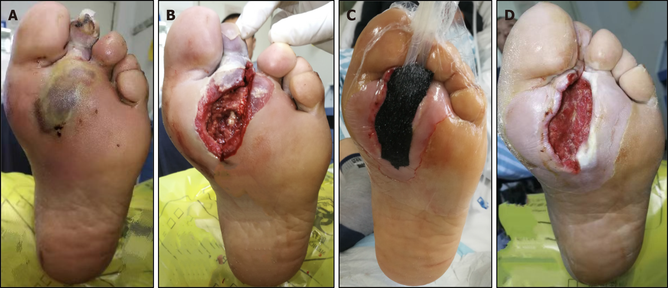
Figure 1 A male patient with diabetic foot ulcer aged 55 years with deep abscess on the left foot for 14 days.
A: Before treatment; B: After debridement surgery; C: Negative pressure wound therapy (NPWT) on the plantar of the foot; D: 7 days after NPWT.
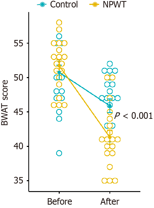
Figure 2 Comparison of Bates-Jensen wound assessment tool scores between the two groups.
BWAT: Bates-Jensen wound assessment tool; NPWT: Negative pressure wound therapy.
Figure 3 Comparison of cluster of differentiation 31 between the two groups.
Cluster of differentiation 31 (CD31) immunohistochemical staining was used, where the brownish yellow or brown color indicates the positive area (40 ×). NPWT: Negative pressure wound therapy.
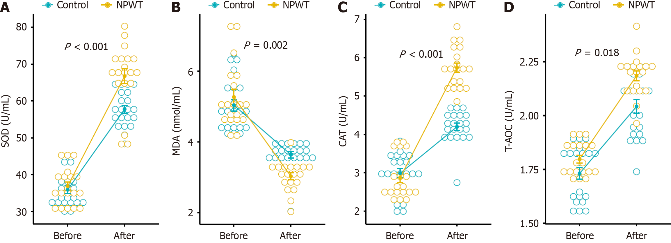
Figure 4 Comparison of levels of oxidative stress makers between the two groups.
A: Superoxide dismutase; B: Malondialdehyde; C: Catalase; D: Total antioxidant capacity. AOC: Total antioxidant capacity; CAT: Catalase; MDA: Malondialdehyde; SOD: Superoxide dismutase; T-NPWT: Negative pressure wound therapy.
Figure 5 Comparison of levels of nuclear factor erythroid 2-related factor 2 and Kelch-like epichlorohydrin-associated protein 1 between the two groups.
aP < 0.05. Keap1: Kelch-like epichlorohydrin-associated protein 1; Nrf2: Nuclear factor erythroid 2-related factor 2; NPWT: Negative pressure wound therapy.
- Citation: Sun HJ, Si SW, Ma YM, Liu XK, Geng HF, Liang J. Role of nuclear factor erythroid 2-related factor 2 in negative pressure wound therapy for diabetic foot ulcers. World J Diabetes 2025; 16(5): 104350
- URL: https://www.wjgnet.com/1948-9358/full/v16/i5/104350.htm
- DOI: https://dx.doi.org/10.4239/wjd.v16.i5.104350


