Copyright
©The Author(s) 2023.
World J Diabetes. Jun 15, 2023; 14(6): 820-845
Published online Jun 15, 2023. doi: 10.4239/wjd.v14.i6.820
Published online Jun 15, 2023. doi: 10.4239/wjd.v14.i6.820
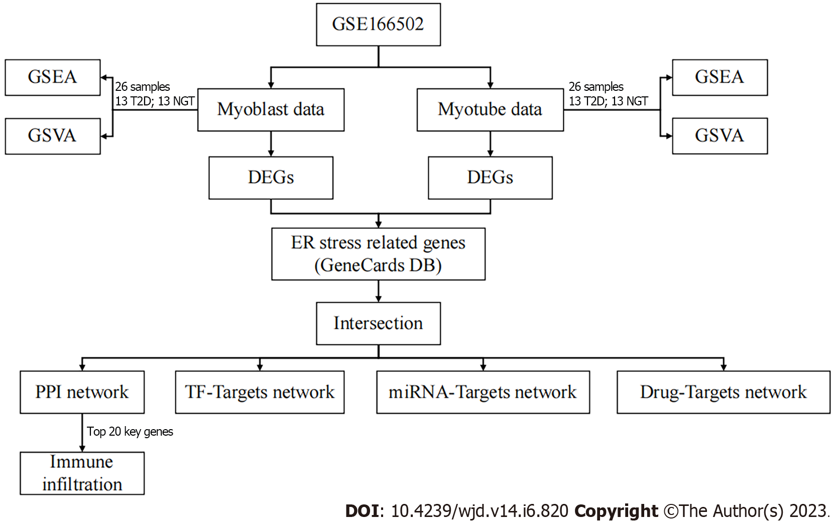
Figure 1 Study flow chart.
GSEA: Gene set enrichment analysis; GSVA: Gene set variation analysis; ER: Endoplasmic reticulum; DEG: Differentially expressed genes; PPI: Protein–protein interaction; TF: Transcription factor.
Figure 2 Gene set enrichment analysis.
A: Top five gene set enrichment analysis in proliferating myoblasts; B: Neuroactive ligand–receptor interaction; C: Hypertrophic cardiomyopathy; D: DNA replication; E: Cell cycle; F: Cardiac muscle contraction; G: Bubble plot in proliferating myoblasts; H: Ridgeline plot in proliferating myoblasts; I: Top 5 gene set enrichment analysis in differentiated myotubes; J: Viral myocarditis; K: Steroid hormone biosynthesis; L: Hematopoietic cell lineage; M: Focal adhesion; N: Extracellular matrix–receptor interaction; O: Bubble plot in differentiated myotubes; P: Ridgeline plot in differentiated myotubes.
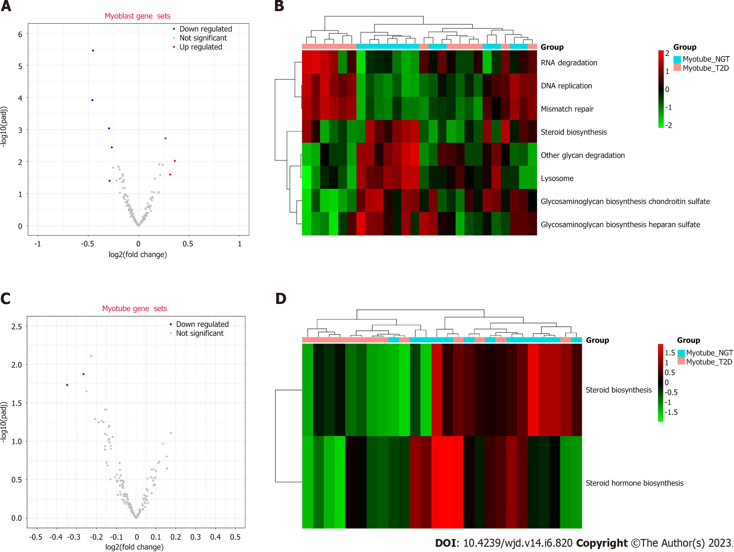
Figure 3 Gene set variation analysis.
A: Volcano plot of proliferating myoblasts; B: Volcano plot of differentiated myotubes; C: Heat map of proliferating myoblasts; D: Heat map of differentiated myotubes.
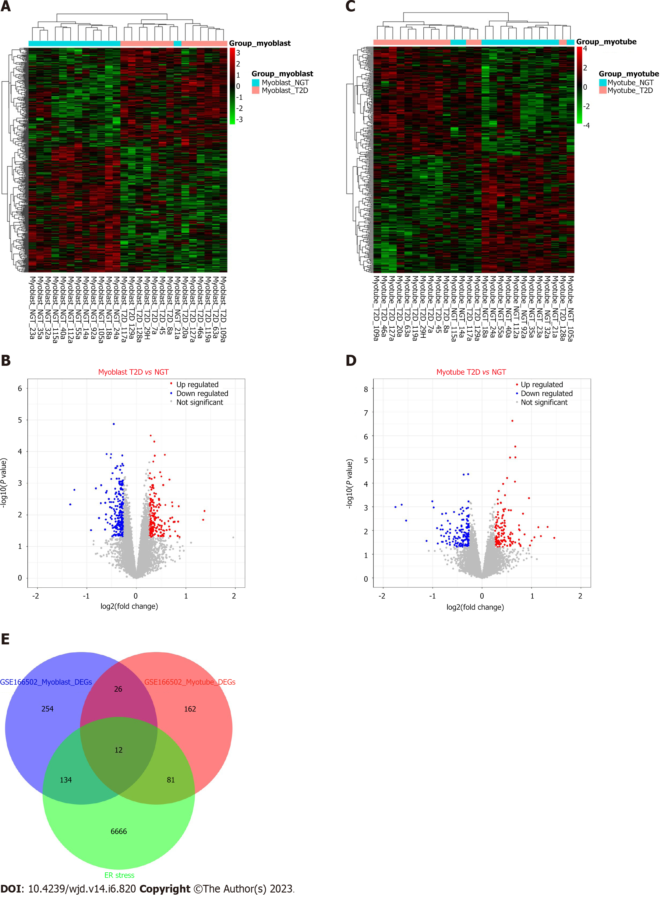
Figure 4 Endoplasmic reticulum stress-related differentially expressed genes.
A: Heat map of proliferating myoblasts; B: Volcano plot of proliferating myoblasts; C: Heat map of differentiated myotubes; D: Volcano plot of differentiated myotubes; E: Endoplasmic reticulum stress-related differentially expressed genes. ER: Endoplasmic reticulum.
Figure 5 Functional enrichment analysis.
A: Gene Ontogeny biological processes; B: Gene Ontogeny cellular components; C: Gene Ontogeny molecular function; D: Kyoto Encyclopedia of Genes and Genomes.
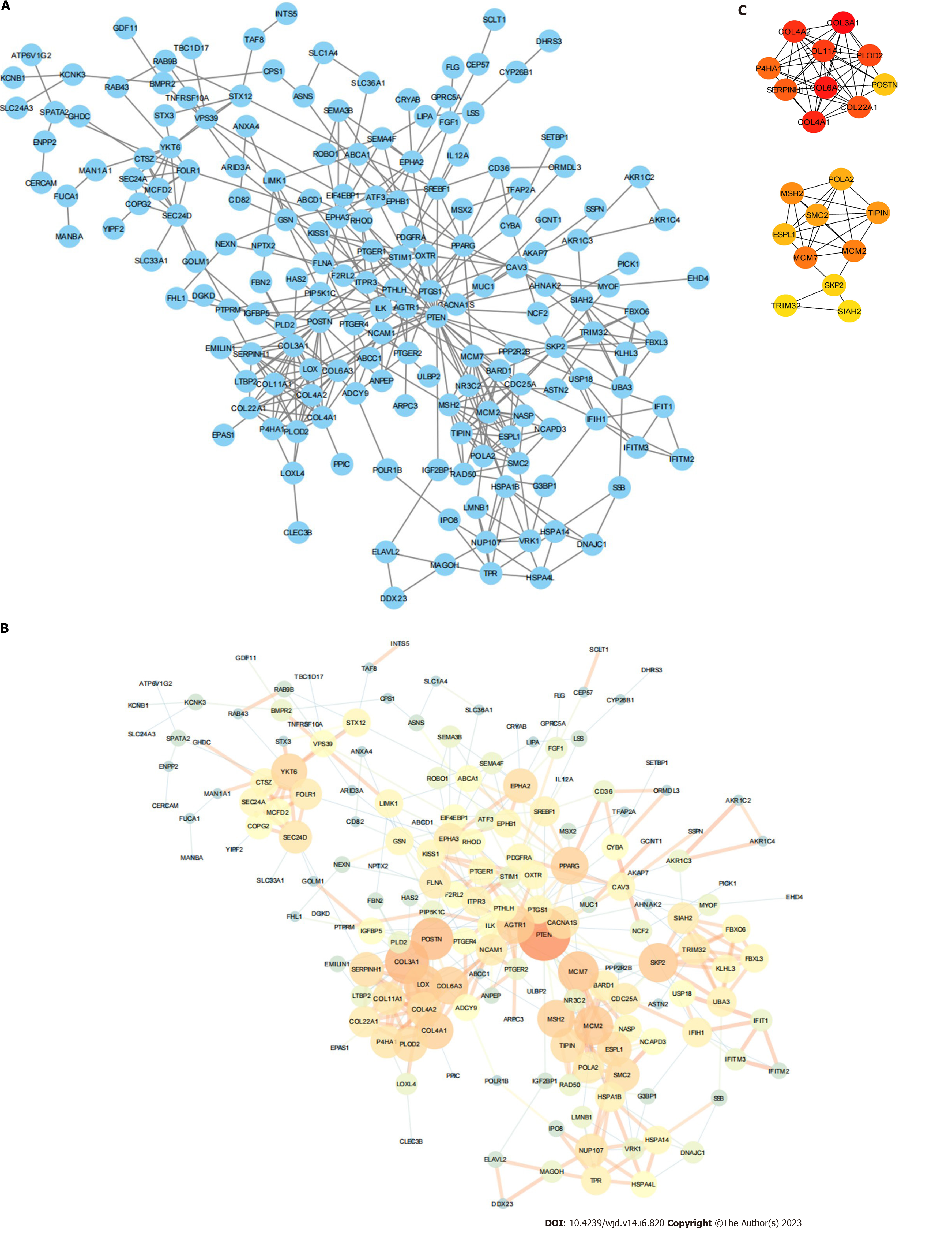
Figure 6 Protein–protein interaction network.
A: Protein–protein interaction (PPI) network; B: PPI network by NetworkAnalyzer; C: PPI network of 20 key genes.
Figure 7 Networks.
A: Transcription factor–mRNA network; B: miRNA–mRNA network; C: Drug–mRNA network.
Figure 8 Immune infiltration.
A: Histogram of immune infiltration distribution; B: Histogram of immune infiltration sample distribution; C: Heat map of immune infiltration correlation; D: Correlation diagram of 20 key genes.
- Citation: Liang B, Chen SW, Li YY, Zhang SX, Zhang Y. Comprehensive analysis of endoplasmic reticulum stress-related mechanisms in type 2 diabetes mellitus. World J Diabetes 2023; 14(6): 820-845
- URL: https://www.wjgnet.com/1948-9358/full/v14/i6/820.htm
- DOI: https://dx.doi.org/10.4239/wjd.v14.i6.820


