Copyright
©The Author(s) 2023.
World J Diabetes. Jan 15, 2023; 14(1): 35-47
Published online Jan 15, 2023. doi: 10.4239/wjd.v14.i1.35
Published online Jan 15, 2023. doi: 10.4239/wjd.v14.i1.35
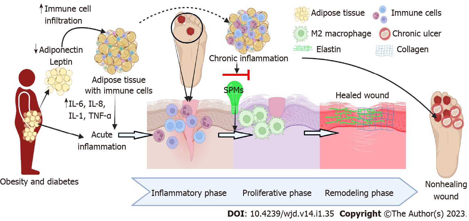
Figure 1 Inflammation-mediated pathogenesis of diabetic foot ulcer, the role of resolvins, and phases of wound healing.
Resolvins [specialized pro-resolving mediators (SPMs)] facilitate the resolution of inflammation and progression of the wound to the resolution phase followed by remodeling and healing (SPMs shown in green). However, persistent infiltration of immune cells and increased secretion of cytokines mediate chronic inflammation and hold the wound in the inflammation phase without progressing to resolution or proliferative phase (red arrow). This leads to the chronicity of inflammation and nonhealing of diabetic foot ulcers. ILV: Intraluminal vesicle.
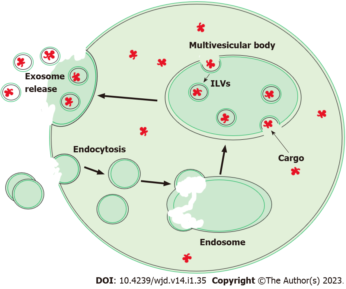
Figure 2 Exosome formation through the endosomal pathway.
Endocytosis produces endocytic vesicles which will fuse to form early endosomes. Endosomes mature into multivesicular bodies (MVBs) and parts of their membranes endocytose to form intraluminal vesicles (ILVs) within themselves. With 2 stages of endocytosis, the orientation of the bilaminar membrane of the ILVs will possess the same orientation as the cell’s membrane. The MVBs fuse to the cellular membrane to release the ILVs now referred to as exosomes. IL: Interleukin; TNF: Tumor necrosis factor.
- Citation: Littig JPB, Moellmer R, Agrawal DK, Rai V. Future applications of exosomes delivering resolvins and cytokines in facilitating diabetic foot ulcer healing. World J Diabetes 2023; 14(1): 35-47
- URL: https://www.wjgnet.com/1948-9358/full/v14/i1/35.htm
- DOI: https://dx.doi.org/10.4239/wjd.v14.i1.35


