Published online Nov 15, 2017. doi: 10.4251/wjgo.v9.i11.452
Peer-review started: June 1, 2017
First decision: July 10, 2017
Revised: July 19, 2017
Accepted: August 17, 2017
Article in press: August 17, 2017
Published online: November 15, 2017
Processing time: 173 Days and 17 Hours
We describe an uncommon case of a patient with a metastatic adenocarcinoma of ovarian origin presented as a gastric subepithelial tumor (SET) and that was diagnosed by endoscopic ultrasound fine-needle biopsy (EUS-FNB). Malignant gastric lesions are rarely metastatic and the primary tumor is mainly breast, lung, esophageal cancer or cutaneous melanoma. Gastric metastasis from ovarian cancer is unusual, presenting synchronously with the primary tumor but also several years later than the initial diagnosis. From an endoscopic point of view, gastric metastasis does not present specific features. They may mimic both a primary gastric tumor or, less frequently, an SET. This case demonstrates the importance of EUS-FNB in distinguishing SETs and how this may alter treatment and prognosis.
Core tip: Gastric metastasis from ovarian cancer is unusual, either as synchronous with the primary tumor or appearing several years after its initial diagnosis. The diagnosis is challenging because of the low incidence, especially when gastric metastases present as a subepithelial tumor. This case emphasizes the crucial role of endoscopic ultrasound fine-needle biopsy in the differential diagnosis of this rare condition.
- Citation: Antonini F, Laterza L, Fuccio L, Marcellini M, Angelelli L, Calcina S, Rubini C, Macarri G. Gastric metastasis from ovarian adenocarcinoma presenting as a subepithelial tumor and diagnosed by endoscopic ultrasound-guided tissue acquisition. World J Gastrointest Oncol 2017; 9(11): 452-456
- URL: https://www.wjgnet.com/1948-5204/full/v9/i11/452.htm
- DOI: https://dx.doi.org/10.4251/wjgo.v9.i11.452
Gastrointestinal (GI) subepithelial tumors (SETs) are lesions located under a normal-appearing mucosa that include several neoplastic and non-neoplastic conditions. Most of the GI-SETs are asymptomatic, therefore their real incidence is unknown. Stomach is the GI tract where the highest incidence is documented[1]. The diagnosis of SETs can be challenging because conventional endoscopic biopsies are frequently inconclusive. Endoscopic ultrasound (EUS) is currently recommended as the preferred investigation modality to establish the exact nature of SETs because of its accuracy in differentiating them from extrinsic compression and providing information about morphology and layer of origin[2]. Lesions arising from the muscularis propria usually represent mesenchymal tumors, such as gastrointestinal stromal tumors (GIST), leiomyomas and schwannomas[1,2]. Metastasis to the GI tract generally involves the fourth and fifth layers and can be misleading as a GIST[1,2].
Herein, we describe an uncommon case of a patient with a metastatic adenocarcinoma of ovarian origin presented as a gastric SET mimicking a GIST and diagnosed by EUS-guided tissue acquisition.
The patient was a 61-year-old woman diagnosed in November 2013 with stage IV high-grade serous carcinoma of the ovary treated with cycles of carboplatin and taxol chemotherapy with partial clinical response. After 1-year of therapy, due to the persistence of abdominal and pelvic disease, cytoreductive surgery had been performed. In June 2015, her CA125 levels had increased to 138 U/mL (normal value < 35 U/mL) and a computed tomography (CT) of the abdomen showed progression of the disease.
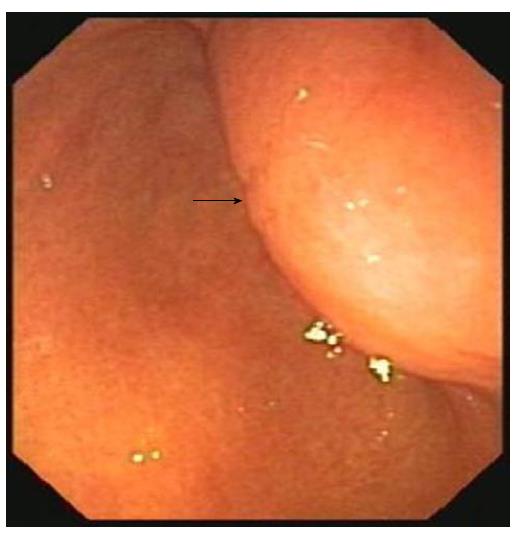
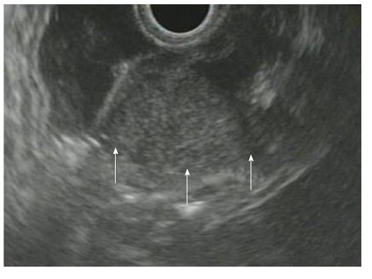
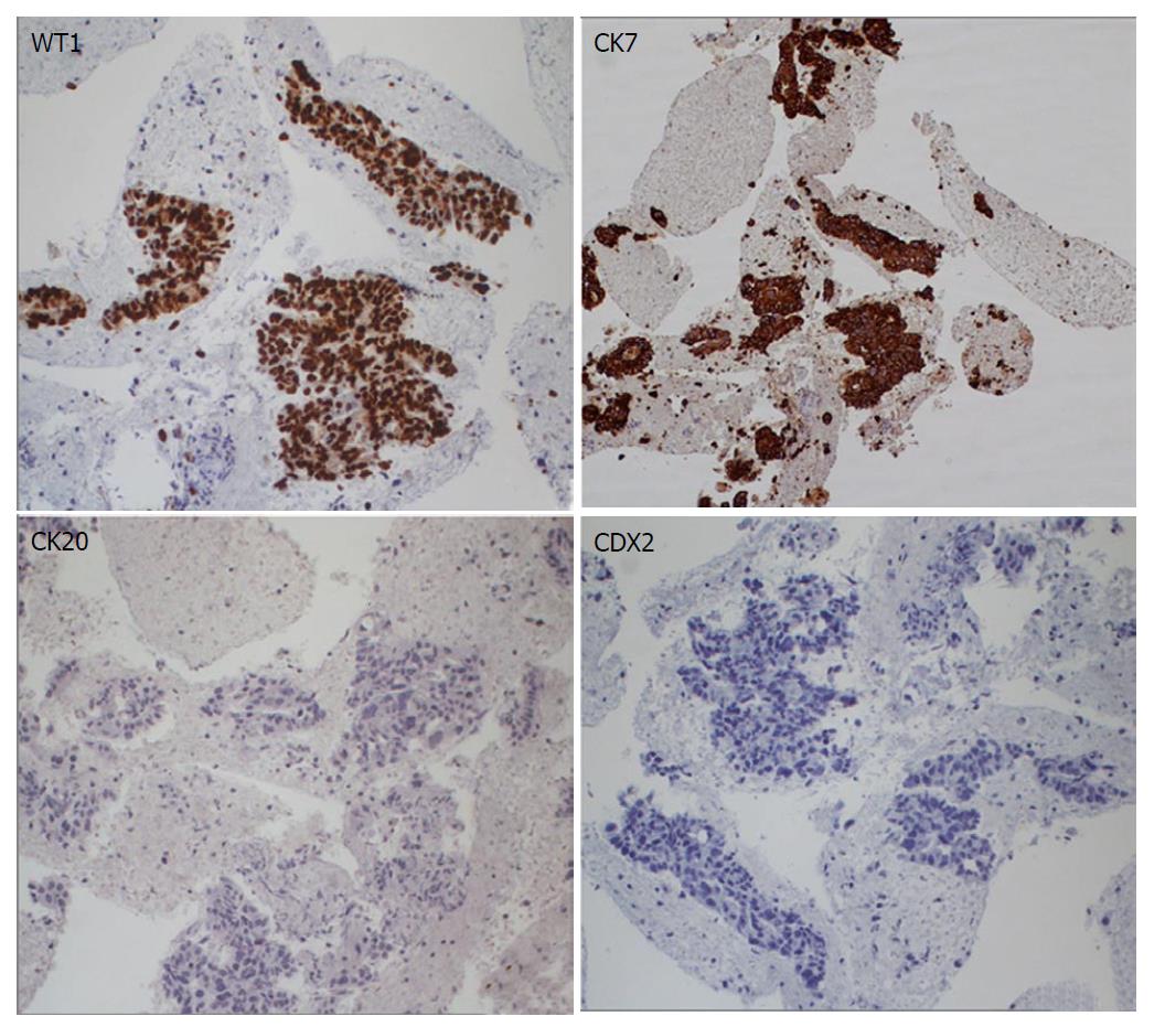
She was referred at that time to our endoscopic center for evaluation of dyspepsia. Upper GI endoscopy revealed an SET covered by normal mucosa on the posterior wall of the gastric antrum (Figure 1). Biopsies of the overlying mucosa proved inconclusive. EUS showed a 23-mm mass within the muscularis propria, hypoechoic but more hyperechoic than the muscular tissue (Figure 2). EUS-guided fine needle biopsy (FNB) was performed for tissue diagnosis. Histology showed adenocarcinoma with immunohistochemistry positive for WT1 and CK7, and negative for CK20 and CDX2 (Figure 3). These findings supported the final diagnosis of a metastatic adenocarcinoma of ovarian origin. The patient started paclitaxel chemotherapy and was alive at the 18-mo follow-up visit.
Gastric metastasis is rare and has been reported mainly from breast, lung, esophageal cancer or cutaneous melanoma[3]. Ovarian carcinoma regularly metastasizes to the peritoneal surface[4]. The acquisition of invasiveness in ovarian carcinoma is accompanied by the process of epithelial to mesenchymal transition. Cancer-associated fibroblasts originating from stromal fibroblastic cells are a component of the tumor microenvironment and promote tumor angiogenesis and lymphangiogenesis[5]. Also, the MUC4 mucin has a role in the invasiveness of ovarian cancer cells because it is overexpressed in ovarian tumors. The overexpression of MUC4 in ovarian cancer is a morphological alteration, along with a decreased expression of epithelial markers (E-cadherin and cytokeratin-18) and an increased expression of mesenchymal markers (N-cadherin and vimentin)[6].
GI involvement from ovarian cancer is limited to the seromuscular layer of the small and large bowels but it also metastasizes through the lymphatic and hematogenous route, with a frequency ranging from 0.7% to 1.8%[7]. The stomach is highly vascularized, therefore the dissemination of ovarian carcinoma is possible but rare. Gastric metastasis from ovarian cancer is unusual, either as synchronous with the primary tumor or appearing several years after its initial diagnosis[8]. The diagnosis is challenging because of its low incidence. Clinical manifestations include epigastric pain, nausea, vomiting, anemia, melena or occult GI blood loss. Obstructive symptoms may be present in case of involvement of the cardia or pylorus[9]. In asymptomatic patients, CA-125 levels beyond normal range may be the only warning sign[7,9]. The prognosis of gastric metastases of ovarian carcinoma is still unknown, a 1-year survival rate can be optimistically expected[10,11]. From an endoscopic point of view, gastric metastases do not present specific features. They may mimic both a primary gastric tumor or, less frequently, an SET[8,12] and can be solitary or more rarely multiple[13].
Several cases of metastatic ovarian cancer presenting as gastric SET have been reported in the literature[10,13-17] but only few have been diagnosed by EUS-guided tissue acquisition, as in our case (Table 1). In other cases, surgical excision or endoscopic submucosal dissection with enucleation has been performed[8,11].
| Ref. | Age | Clinicalpresentation | Diagnosis of ovarian cancer | Endoscopicaspect of gastric SET | EUS morphology |
| Shanga et al[13], 2003 | 62 | Epigastric discomfort | 7 yr before | Body, 4 cm | Irregular border, hypoechoic lesion, fourth layer |
| Jung et al[10], 2009 | 49 | Asymptomatic | 52 mo from surgery | Antrum, 2.5 cm × 2.5 cm | Hypoechoic lesion, fourth layer |
| Carrara et al[14], 2011 | 70 | Mild anemia, dyspepsia | NR | Body, 3.8 cm × 4.8 cm, ulcerated | Third layer |
| Akce et al[15], 2012 | 55 | Anemia, melena | 5 yr before | Antrum, 3.4 cm × 3.7 cm and body, 1.2 cm × 0.8 cm | Hypoechoic lesions, fourth layer |
| Yamao et al[16], 2015 | 51 | NR | 25 mo from surgery | Antrum, 3 cm | Hypoechoic lesion with marginal rim, fourth layer |
| Current case | 61 | Dyspepsia | 2 yr before | Antrum, 2.3 cm | Hypoechoic lesion (more hyperechoic than the muscular tissue), fourth layer |
In the present case, the lesion was mimicking a GIST, even if a metastasis from ovarian cancer had been considered. For these reasons, a tissue diagnosis was considered necessary. After a diagnosis of metastatic ovarian cancer to the stomach had been achieved, surgical intervention or more aggressive options would become unnecessary considering the progression of the disease.
In conclusion, although rare, gastric metastasis from primary ovarian cancer should be considered in any patient with a history of ovarian adenocarcinoma who presents with gastric tumor. This case emphasizes the crucial role of EUS-FNB in the differential diagnosis.
A 61-year-old woman diagnosed 3 years previously with stage IV high-grade serous carcinoma of the ovary and treated with debulking surgery and chemotherapy presented for evaluation of dyspepsia.
General physical examination was unremarkable.
Gastrointestinal stromal tumor, gastric tumor, metastasis.
CA125 levels had increased to 138 U/mL (normal value < 35 U/mL).
Endoscopic ultrasonography (EU) showed a 23-mm mass within the muscularis propria, hypoechoic but more hyperechoic than the muscular tissue.
Histology obtained via EUS-guided fine needle biopsy showed adenocarcinoma with immunohistochemistry positive for WT1 and CK7, and negative for CK20 and CDX2, supporting the final diagnosis of a metastatic adenocarcinoma of ovarian origin.
Palliative chemotherapy.
Several cases of metastatic ovarian cancer presenting as gastric subepithelial tumor have been reported in the literature, but only few of them have been diagnosed by EUS-guided tissue acquisition, as in our case. In other cases, surgical excision or endoscopic submucosal dissection with enucleation has been performed.
Although rare, metastatic ovarian cancer to the stomach should be considered in any patient with a history of ovarian adenocarcinoma who presents with gastric tumor.
This is an interesting case report.
Manuscript source: Invited manuscript
Specialty type: Oncology
Country of origin: Italy
Peer-review report classification
Grade A (Excellent): 0
Grade B (Very good): B
Grade C (Good): C, C
Grade D (Fair): 0
Grade E (Poor): 0
P- Reviewer: Erturk SM, Langdon S, Sergi CM S- Editor: Ji FF L- Editor: A E- Editor: Zhao LM
| 1. | Landi B, Palazzo L. The role of endosonography in submucosal tumours. Best Pract Res Clin Gastroenterol. 2009;23:679-701. [RCA] [PubMed] [DOI] [Full Text] [Cited by in Crossref: 35] [Cited by in RCA: 50] [Article Influence: 3.1] [Reference Citation Analysis (0)] |
| 2. | Papanikolaou IS, Triantafyllou K, Kourikou A, Rösch T. Endoscopic ultrasonography for gastric submucosal lesions. World J Gastrointest Endosc. 2011;3:86-94. [RCA] [PubMed] [DOI] [Full Text] [Full Text (PDF)] [Cited by in CrossRef: 51] [Cited by in RCA: 78] [Article Influence: 5.6] [Reference Citation Analysis (0)] |
| 3. | Campoli PM, Ejima FH, Cardoso DM, Silva OQ, Santana Filho JB, Queiroz Barreto PA, Machado MM, Mota ED, Araujo Filho JA, Alencar Rde C. Metastatic cancer to the stomach. Gastric Cancer. 2006;9:19-25. [RCA] [PubMed] [DOI] [Full Text] [Cited by in Crossref: 55] [Cited by in RCA: 63] [Article Influence: 3.3] [Reference Citation Analysis (0)] |
| 4. | Kang WD, Kim CH, Cho MK, Kim JW, Lee JS, Ryu SY, Kim YH, Choi HS, Kim SM. Primary epithelial ovarian carcinoma with gastric metastasis mimic gastrointestinal stromal tumor. Cancer Res Treat. 2008;40:93-96. [RCA] [PubMed] [DOI] [Full Text] [Cited by in Crossref: 8] [Cited by in RCA: 14] [Article Influence: 0.8] [Reference Citation Analysis (0)] |
| 5. | Wei R, Lv M, Li F, Cheng T, Zhang Z, Jiang G, Zhou Y, Gao R, Wei X, Lou J. Human CAFs promote lymphangiogenesis in ovarian cancer via the Hh-VEGF-C signaling axis. Oncotarget. 2017;8:67315-67328. [RCA] [PubMed] [DOI] [Full Text] [Full Text (PDF)] [Cited by in Crossref: 23] [Cited by in RCA: 37] [Article Influence: 4.6] [Reference Citation Analysis (0)] |
| 6. | Ponnusamy MP, Lakshmanan I, Jain M, Das S, Chakraborty S, Dey P, Batra SK. MUC4 mucin-induced epithelial to mesenchymal transition: a novel mechanism for metastasis of human ovarian cancer cells. Oncogene. 2010;29:5741-5754. [RCA] [PubMed] [DOI] [Full Text] [Full Text (PDF)] [Cited by in Crossref: 101] [Cited by in RCA: 111] [Article Influence: 7.4] [Reference Citation Analysis (0)] |
| 7. | Pernice M, Manci N, Marchetti C, Morano G, Boni T, Bellati F, Panici PB. Solitary gastric recurrence from ovarian carcinoma: a case report and literature review. Surg Oncol. 2006;15:267-270. [RCA] [PubMed] [DOI] [Full Text] [Cited by in Crossref: 5] [Cited by in RCA: 7] [Article Influence: 0.4] [Reference Citation Analysis (0)] |
| 8. | Zhou JJ, Miao XY. Gastric metastasis from ovarian carcinoma: a case report and literature review. World J Gastroenterol. 2012;18:6341-6344. [RCA] [PubMed] [DOI] [Full Text] [Full Text (PDF)] [Cited by in CrossRef: 9] [Cited by in RCA: 17] [Article Influence: 1.3] [Reference Citation Analysis (0)] |
| 9. | Kadakia SC, Parker A, Canales L. Metastatic tumors to the upper gastrointestinal tract: endoscopic experience. Am J Gastroenterol. 1992;87:1418-1423. [PubMed] |
| 10. | Jung HJ, Lee HY, Kim BW, Jung SM, Kim HG, Ji JS, Choi H, Lee BI. Gastric Metastasis from Ovarian Adenocarcinoma Presenting as a Submucosal Tumor without Ulceration. Gut Liver. 2009;3:211-214. [RCA] [PubMed] [DOI] [Full Text] [Full Text (PDF)] [Cited by in Crossref: 13] [Cited by in RCA: 20] [Article Influence: 1.3] [Reference Citation Analysis (0)] |
| 11. | Kim EY, Park CH, Jung ES, Song KY. Gastric metastasis from ovarian cancer presenting as a submucosal tumor: a case report. J Gastric Cancer. 2014;14:138-141. [RCA] [PubMed] [DOI] [Full Text] [Full Text (PDF)] [Cited by in Crossref: 3] [Cited by in RCA: 8] [Article Influence: 0.7] [Reference Citation Analysis (0)] |
| 12. | Oda , Kondo H, Yamao T, Saito D, Ono H, Gotoda T, Yamaguchi H, Yoshida S, Shimoda T. Metastatic tumors to the stomach: analysis of 54 patients diagnosed at endoscopy and 347 autopsy cases. Endoscopy. 2001;33:507-510. [RCA] [PubMed] [DOI] [Full Text] [Cited by in Crossref: 158] [Cited by in RCA: 165] [Article Influence: 6.9] [Reference Citation Analysis (0)] |
| 13. | Sangha S, Gergeos F, Freter R, Paiva LL, Jacobson BC. Diagnosis of ovarian cancer metastatic to the stomach by EUS-guided FNA. Gastrointest Endosc. 2003;58:933-935. [PubMed] |
| 14. | Carrara S, Doglioni C, Arcidiacono PG, Testoni PA. Gastric metastasis from ovarian carcinoma diagnosed by EUS-FNA biopsy and elastography. Gastrointest Endosc. 2011;74:223-225. [RCA] [PubMed] [DOI] [Full Text] [Cited by in Crossref: 13] [Cited by in RCA: 13] [Article Influence: 0.9] [Reference Citation Analysis (0)] |
| 15. | Akce M, Bihlmeyer S, Catanzaro A. Multiple gastric metastases from ovarian carcinoma diagnosed by endoscopic ultrasound with fine needle aspiration. Case Rep Gastrointest Med. 2012;2012:610527. [RCA] [PubMed] [DOI] [Full Text] [Full Text (PDF)] [Cited by in Crossref: 1] [Cited by in RCA: 10] [Article Influence: 0.8] [Reference Citation Analysis (0)] |
| 16. | Yamao K, Kitano M, Kudo M, Maenishi O. Synchronous pancreatic and gastric metastasis from an ovarian adenocarcinoma diagnosed by endoscopic ultrasound-guided fine-needle aspiration. Endoscopy. 2015;47 Suppl 1 UCTN:E596-E597. [RCA] [PubMed] [DOI] [Full Text] [Cited by in Crossref: 2] [Cited by in RCA: 5] [Article Influence: 0.6] [Reference Citation Analysis (0)] |
| 17. | Liu Q, Yu QQ, Wu H, Zhang ZH, Guo RH. Isolated gastric recurrence from ovarian carcinoma: A case report. Oncol Lett. 2015;9:1173-1176. [RCA] [PubMed] [DOI] [Full Text] [Full Text (PDF)] [Cited by in Crossref: 2] [Cited by in RCA: 4] [Article Influence: 0.4] [Reference Citation Analysis (0)] |


