Published online Dec 15, 2010. doi: 10.4251/wjgo.v2.i12.443
Revised: November 14, 2010
Accepted: November 21, 2010
Published online: December 15, 2010
Isolated extragonadal germ cell tumors can be primary in nature or metastatic from a burned out testicular cancer. Accurate diagnosis is critical as appropriate therapy can be highly curative. We present the case of an isolated extragonadal germ cell tumor in the retroperitoneum diagnosed by endoscopic ultrasound-guided fine needle aspiration. This case underscores the importance of considering germ cell tumors in the differential diagnosis of an unexplained retroperitoneal mass, particularly since immunophenotypic staining may be necessary to establish the diagnosis.
- Citation: Womeldorph CM, Zalupski MM, Knoepp SM, Soltani M, Elmunzer BJ. Retroperitoneal germ cell tumor diagnosed by endoscopic ultrasound-guided fine needle aspiration. World J Gastrointest Oncol 2010; 2(12): 443-445
- URL: https://www.wjgnet.com/1948-5204/full/v2/i12/443.htm
- DOI: https://dx.doi.org/10.4251/wjgo.v2.i12.443
Germ cell neoplasms are relatively uncommon but highly curable when recognized and treated appropriately[1]. They most commonly present as a palpable testicular mass and have a tendency to metastasize to nodal tissue in the retroperitoneum[1]. Extragonadal germ cell neoplasms, either primary or metastatic, can occur in the anterior mediastinum, retroperitoneum and the pineal or suprasellar regions[2]. Bulky lesions generally cause clinical symptoms including chest pain, dyspnea, cough, abdominal pain and back pain, depending on their location[3]. In this report, we present an interesting case of an asymptomatic man with an isolated retroperitoneal germ cell tumor that was diagnosed by endoscopic ultrasound-guided fine needle aspiration (EUS-FNA).
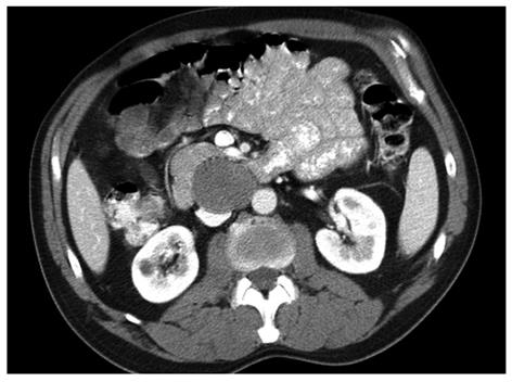
A 55-year old asymptomatic man presented to the emergency room after undergoing a whole body “health screening”computed tomography (CT) scan that revealed a large retroperitoneal abnormality, concerning for a dissecting aortic aneurysm. A repeat contrasted CT scan of the abdomen revealed a 4.7 cm × 4.3 cm mass in the region of the 3rd portion of the duodenum with mass effect on the inferior vena cava (IVC) (Figure 1). Baseline labs were unremarkable, including a normal CA 19-9.
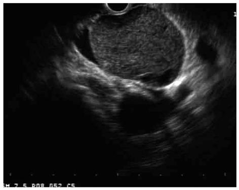
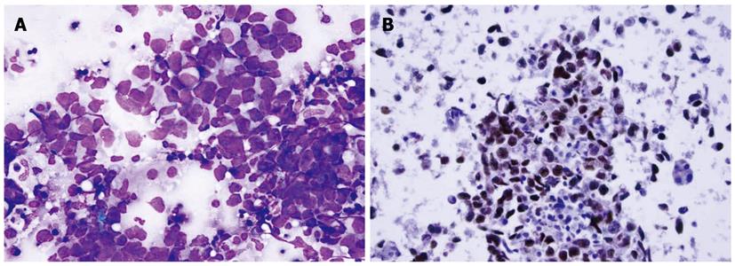
EUS with a radial array echoendoscope (GF-UE160-AL5, Olympus Medical Systems Corp, Center Valley, PA, USA) revealed a 4 cm × 3 cm well-circumscribed retroperitoneal mass abutting the IVC, aorta and uncinate process of the pancreas (Figure 2). FNA with a 22-gauge needle (EchoTip 1-22, Cook Medical, Bloomington, IN, USA) through a curved linear array echoendoscope (GF-UC140P-AL5, Olympus) yielded a preliminary cytological diagnosis of undifferentiated anaplastic carcinoma of unclear origin (Figure 3A). After discussion at multidisciplinary tumor board, germ cell-specific immunophenotypic staining was requested and demonstrated that the tumor strongly expressed OCT4 and PLAP, weakly expressed c-kit and was negative for AFP, HCG and CD30, a pattern consistent with metastatic germ cell tumor, favoring seminoma (Figure 3B).
Testicular examination was notable for an atrophic right testicle without mass. Testicular ultrasound revealed a small, heterogeneous right testicle with multiple hypoechoic areas but no definite mass. Subsequent right inguinal orchiectomy and pathological evaluation revealed a focal testicular parenchymal scar consistent with spontaneous regression of a burned out germ cell tumor. The retroperitoneal mass was treated with radiation therapy with curative intent.
Endoscopic ultrasound was originally developed for accurate evaluation of the pancreas. With time, it became apparent that EUS was also effective in the staging of a multitude of gastrointestinal cancers as well as the characterization of subepithelial lesions of the GI tract. The development of the curved linear array echoendoscope allowed sonographically-guided fine needle aspiration, making EUS one of the most important tools in the diagnosis of digestive tract tumors. EUS-FNA is also very accurate in the diagnosis of non-digestive lesions adjacent to the GI tract[4,5]. Diagnostic tissue can be obtained from a multitude of non-gastrointestinal lesions, benign or malignant. To our knowledge, this is the first case of an isolated germ cell tumor involving the retroperitoneum diagnosed by EUS-FNA.
Isolated extragonadal germ cell tumors may be metastatic from a testicular primary that has regressed or primary due to malignant transformation of germ cells that have mismigrated along the urogenital ridge during embryogenesis[3,6]. On this basis, all extragonadal germ cell tumors in men should prompt physical and sonographic examination of the testes to exclude a silent or burned out primary cancer[7,8]. Furthermore, because specialized immunophenotypic testing is necessary to accurately categorize these lesions, this case underscores the importance of considering an extragonadal germ cell tumor in the differential diagnosis of unexplained retroperitoneal masses, especially those with poorly differentiated or anaplastic histology.
Peer reviewer: Akihiko Tsuchida, MD, PhD, Associate Professor, Department of Surgery, Tokyo Medical University, 6-7-1 Nishi-Shinjuku, Shinjuku-ku, Tokyo 160-0023, Japan
S- Editor Wang JL L- Editor Roemmele A E- Editor Ma WH
| 1. | Collins DH, Pugh RC. Classification and frequency of testicular tumours. Br J Urol. 1964;36:SUPPL: 1-11. |
| 2. | Richie JP. Neoplasms of the testis. Campbell’s urology. 6th ed. Philadelphia: W. B. Saunders Co 1992; 1222-1263. |
| 3. | Bokemeyer C, Nichols CR, Droz JP, Schmoll HJ, Horwich A, Gerl A, Fossa SD, Beyer J, Pont J, Kanz L. Extragonadal germ cell tumors of the mediastinum and retroperitoneum: results from an international analysis. J Clin Oncol. 2002;20:1864-1873. |
| 4. | Anand D, Barroeta JE, Gupta PK, Kochman M, Baloch ZW. Endoscopic ultrasound guided fine needle aspiration of non-pancreatic lesions: an institutional experience. J Clin Pathol. 2007;60:1254-1262. |
| 5. | Bentz JS, Kochman ML, Faigel DO, Ginsberg GG, Smith DB, Gupta PK. Endoscopic ultrasound-guided real-time fine-needle aspiration: clinicopathologic features of 60 patients. Diagn Cytopathol. 1998;18:98-109. |
| 6. | Willis RA. Borderland of embryology and pathology. Washington, DC: Butterworth and Co 1962; 442. |


