Published online Oct 15, 2010. doi: 10.4251/wjgo.v2.i10.395
Revised: August 20, 2010
Accepted: August 27, 2010
Published online: October 15, 2010
The diagnosis of gastric metastasis from lung cancer is relatively rare in living patients. We describe a case of Type 4 tumor-like metastasis due to primary lung cancer diagnosed with immunohistochemical staining while the patient was alive. A 68-year-old man was admitted to our hospital because of epigastric pain. Gastrointestinal endoscopy revealed a Type 4 tumor and the histological examination showed poorly differentiated adenocarcinoma. His chest X-ray showed mass shadow in the right upper lung field. The resected specimens showed moderately differentiated adenocarcinoma., The diagnosis of gastric metastasis from lung cancer was made by immunohistochemical staining of the lung and gastric tumors which showed positive staining for Thyroid transcriptional factor-1. Diagnosis of gastric metastasis, especially Type 4 metastasis by lung cancer is difficult. However, immunohistochemical staining is very helpful for diagnosis of primary lung cancer metastasis at sites such as the gastrointestinal tract which are not normally prone to metastatis.
- Citation: Okazaki R, Ohtani H, Takeda K, Sumikawa T, Yamasaki A, Matsumoto S, Shimizu E. Gastric metastasis by primary lung adenocarcinoma. World J Gastrointest Oncol 2010; 2(10): 395-398
- URL: https://www.wjgnet.com/1948-5204/full/v2/i10/395.htm
- DOI: https://dx.doi.org/10.4251/wjgo.v2.i10.395
Gastric metastasis from lung cancer is relatively rare with a reported incidence of 0.2% to 0.5%[1,2] at necropsy. In fact, the diagnosis of gastric metastasis due to primary lung cancer is very rare in living patients. The macroscopic findings of gastric metastasis in most case reveal submucosal tumor formation radiographically demonstrated as a Bull’s eye sign[3] while Type 4 tumor-like metastasis is relatively rare. Here, we describe a case of gastric metastasis showing Type 4 tumor-like metastasis due to primary lung cancer diagnosed by immunohistochemical staining.
A 68-year-old man with a 40-year smoking history was admitted to a local hospital because of severe epigastric pain. Gastrointestinal endoscopy was performed and an erosive, an easy-bleeding tumor spread from the upper third of the stomach to the body was found. His chest X-ray film also showed mass shadow in the right lower field. Therefore, the patient was transferred to our hospital for the further examination.
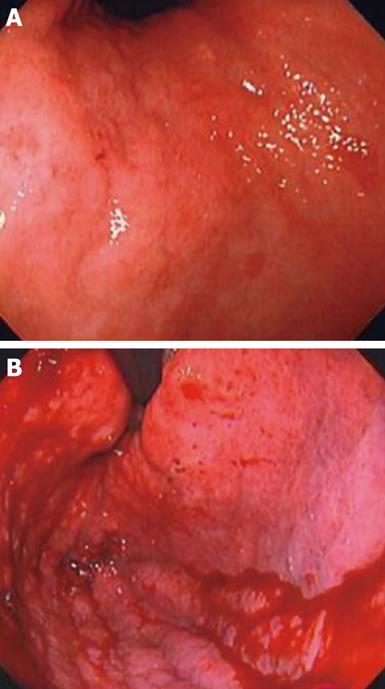
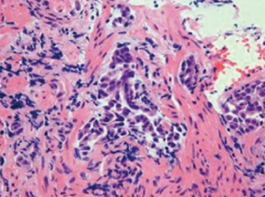
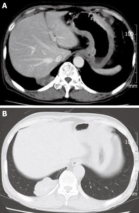
Serum tumor markers were markedly elevated (CEA: 294.0 ng/mL, SLX: 440 U/mL, CYFRA: 10 ng/mL, CA19-9: 5260U/mL). Routine blood hematology and chemistries were not remarkable. The abdomen appeared normal on physical examination. However, we found a Type 4 tumor using gastrointestinal endoscopy (Figure 1) and the biopsy from the tumor indicated a poorly differentiated adenocarcinoma (Figure 2). A chest computed tomography showed a mass shadow in the right lung S10 segment and thickening of the gastric wall (Figure 3). Transbronchial biopsy of the right S10 tumor was performed and pathological examination of a specimen from the tumor showed adenocarcinoma. Possible diagnoses were metastatic lung cancer from primary gastric cancer, primary lung cancer with gastric metastasis or concomitance of primary lung cancer and primary gastric cancer.
After the transbronchial biopsy, the patient developed lung abscess in the right lower lobe and a right lower lobectomy was performed as the administration of antibiotics was insufficient. Histological examination revealed moderately differentiated adenocarcinoma of the lung. To examine whether the gastric tumor was a primary gastric cancer or a metastasis from the lung cancer, immunohistochemical staining was performed. The diagnosis of gastric metastasis from lung cancer was confirmed by positive staining for Cytokeratin 7 (Figure 4A), Thyroid transcriptional factor-1 (TTF-1) (Figure 4B), Cytokeratin AE 1/3, and CEA.
The patient was treated with three courses of systemic chemotherapy with carboplatin (180 mg/body, days 1, 8 and 15) and paclitaxel (80 mg/m2, days 1, 8 and 15). However, the patient’s general condition gradually worsened because of the progressive metastasis to brain and bone and he died of sepsis one year after the diagnosis of gastric metastasis. An autopsy was not performed.
Metastasis to sites in the gastrointestinal tract are rare[2]. Menuck et al[4] reported that the incidence of gastric metastasis was 1.7% in 1010 autopsy cases and that the most common cancers metastasizing to the stomach were breast cancer, melanoma and lung cancer. On the other hand, primary lung cancer is prone to metastasize to the liver, adrenals, bone, and brain[2,5]. The percentage of gastric metastasis from lung has been reported as 0.2% to 0.5% at necropsy[1,2]. Furthermore, the diagnosis of gastric cancer is very rare in living patients[6].
In the present case, the patient manifested severe epigastric pain and we found gastric metastasis using gastrointestinal endoscopy. However, in most cases, gastric metastases are asymptomatic because they initially occur in the submucosal layer. When the metastatic lesions grow and cause obstruction or ulceration, patients develop symptoms such as abdominal pain, vomiting and anemia. Severe complications due to primary lung cancer, such as gastric perforations, pyloric obstruction have been reported[6-8]. Therefore, when symptoms related to the gastrointestinal tract are present, gastrointestinal endoscopy might be considered although the incidence of gastric metastasis is not high.
In the present case, the gastrointestinal endoscopic findings showed a Type 4 tumor or linitis plastica-like tumor which might be extend as circumscribed plaques within the submucosal layer. Menuck et. al. have described several patterns observed from radiographic findings: (1) solitary polypoid submucosal mass, which may ulcerate; (2) multiple polypoid submucosal masses, which may ulcerate; and (3) infiltrating constricting pattern similar to a “linitis plastica”[4]. Type 4 gastric tumors or linitis plastica-like metastasis are sometimes observed in gastric metastases of breast cancer[9,10]. This pattern of metastasis is very rare in lung cancer and only a few cases have been reported[4,9].
We performed immunohistochemical staining for TTF-1 on tissue sections of gastric adenocarcinoma. TTF-1 is a transcriptional factor and mainly expressed in thyroid and lung during embryogenesis[11]. However, Holtinger et al[12] have reported the usefulness of TTF-1 for diagnosis of primary lung cancer. TTF-1 is immunostained in lung and thyroid cancer but not in breast cancer[13], colon cancer[12] or gastric cancer[14]. Therefore, TTF-1 is recognized as a specific marker for primary lung cancer[15]. In the present case, the diagnosis of metastatic gastric cancer from the endoscopic findings was difficult. However, immunohistochemical staining for TTF-1 was helpful diagnosing lung as the primary site.
In summary, we report a case of lung cancer with Type 4 tumor-like metastasis. Immunohistochemical staining for TTF-1 is useful to distinguish primary gastric cancer from metastatic cancer due to primary lung cancer.
Peer reviewer: Atsushi Imagawa, MD, PhD, Department of Gastroenterology, Tsuyama Central Hospital, 1756 Kawasaki Tsuyama-city, Okayama 708-0841, Japan
S- Editor Wang JL L- Editor Hughes D E- Editor Yang C
| 1. | Antler AS, Ough Y, Pitchumoni CS, Davidian M, Thelmo W. Gastrointestinal metastases from malignant tumors of the lung. Cancer. 1982;49:170-172. |
| 2. | Green LK. Hematogenous metastases to the stomach. A review of 67 cases. Cancer. 1990;65:1596-1600. |
| 3. | Rubin SA, Davis M. "Bull's eye" or "target" lesions of the stomach secondary to carcinoma of the lung. Am J Gastroenterol. 1985;80:67-69. |
| 4. | Menuck LS, Amberg JR. Metastatic disease involving the stomach. Am J Dig Dis. 1975;20:903-913. |
| 5. | Auerbach O, Garfinkel L, Parks VR. Histologic type of lung cancer in relation to smoking habits, year of diagnosis and sites of metastases. Chest. 1975;67:382-387. |
| 6. | Suzaki N, Hiraki A, Ueoka H, Aoe M, Takigawa N, Kishino T, Kiura K, Kanehiro A, Tanimoto M, Harada M. Gastric perforation due to metastasis from adenocarcinoma of the lung. Anticancer Res. 2002;22:1209-1212. |
| 7. | Garwood RA, Sawyer MD, Ledesma EJ, Foley E, Claridge JA. A case and review of bowel perforation secondary to metastatic lung cancer. Am Surg. 2005;71:110-116. |
| 8. | Higgins PM. Pyloric obstructions due to a metastatic deposit from carcinoma of the bronchus. Can J Surg. 1962;5:438-441. |
| 9. | Campoli PM, Ejima FH, Cardoso DM, Silva OQ, Santana Filho JB, Queiroz Barreto PA, Machado MM, Mota ED, Araujo Filho JA, Alencar Rde C. Metastatic cancer to the stomach. Gastric Cancer. 2006;9:19-25. |
| 10. | Kawabata K, Watanabe K, Ozaki S, Ando S, Takegawa S, Kojima Y. A case of breast carcinoma with metastasis to the stomach-Immunoshistochemical differentiation between primary and metastatic gastric carcinoma Borrmannn type 4. Jpn J Breast Cancer. 2004;19:162-167. |
| 11. | Kimura S, Hara Y, Pineau T, Fernandez-Salguero P, Fox CH, Ward JM, Gonzalez FJ. The T/ebp null mouse: thyroid-specific enhancer-binding protein is essential for the organogenesis of the thyroid, lung, ventral forebrain, and pituitary. Genes Dev. 1996;10:60-69. |
| 12. | Holzinger A, Dingle S, Bejarano PA, Miller MA, Weaver TE, DiLauro R, Whitsett JA. Monoclonal antibody to thyroid transcription factor-1: production, characterization, and usefulness in tumor diagnosis. Hybridoma. 1996;15:49-53. |
| 13. | Bejarano PA, Baughman RP, Biddinger PW, Miller MA, Fenoglio-Preiser C, al-Kafaji B, Di Lauro R, Whitsett JA. Surfactant proteins and thyroid transcription factor-1 in pulmonary and breast carcinomas. Mod Pathol. 1996;9:445-452. |
| 14. | Merchant SH, Amin MB, Tamboli P, Ro J, Ordóñez NG, Ayala AG, Czerniak BA, Ro JY. Primary signet-ring cell carcinoma of lung: immunohistochemical study and comparison with non-pulmonary signet-ring cell carcinomas. Am J Surg Pathol. 2001;25:1515-1519. |
| 15. | Sheppard MN. Specific markers for pulmonary tumours. Histopathology. 2000;36:273-276. |


